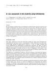
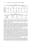
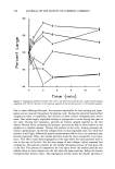
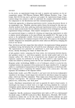
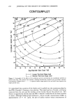
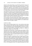
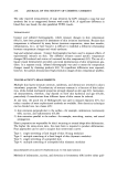
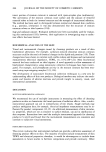
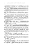
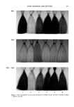
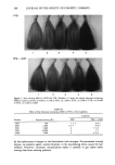
INSTRUMENTAL ASSESSMENT OF SKIN 261 from skin replicas. Images of shadows can be quantitatively analyzed, and results are directly related to surface relief (95,96). Image analysis techniques require specialized equipment: image digitizer (TV camera), video processor, computer, and software. More researchers are using image analysis for skin surface evaluation as complete systems become commercially available (91,97- 100). Recently developed optical instruments (Scopeman, Microwatcher, Nikon) collect and store electronic images directly from the skin surface (101). High-quality images can be evaluated by expert graders and quantified by image analysis techniques (Figure 7). Various magnification lenses are provided to optimize the resolution of skin surface topography however, the depth of field is limited. Scanning electron microscopy provides microscopic images of skin surface replicas with greater depth of field than light microscopy. The images are suitable for subjective and objective evaluations (13,17,82,88,102). These images can be quantified using image analysis techniques. Soap exposure. Quantification of skin surface topography for measuring soap and surfac- tant effects is a new area. Research to date indicates that surface roughness increases after soap exposure. The use of image analysis provides multiple ways to quantify changes in skin microtopography (97,103) however, no standard image analysis technique cur- rently exists. THICKNESS MEASUREMENTS Skin thickness measurements are useful to characterize skin disorders, e.g., psoriasis and eczema, and skin conditions, e.g., edema and epidermal hyperplasia. Ultrasound is the commonly used technique for measuring skin thickness. Surfactant exposure. Ultrasound thickness measurements were conducted after patch tests (Finn chambers) with SLS. Higher concentrations resulted in increased skin thickness as well as increased visual irritation, TEWL, and LDV (27). Figure 7. a. Scopeman for documenung images of skin at various magnifications. b. Image collected by Scopeman.

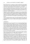
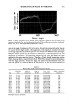
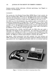
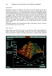
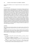
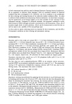
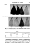
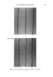
262 JOURNAL OF THE SOCIETY OF COSMETIC CHEMISTS There are no reports to indicate changes in skin thickness after soap exposure under actual use conditions however, surfactant may induce epidermal hyperplasia (104). BIOLOGICAL METHODS Quantitative methods exist to measure skin structures and physiological changes caused by application of topical products. The goal of noninvasive biological assessment is to directly measure the condition of the viable epidermal layers. EXFOLIATIVE CYTOLOGY Noninvasive techniques are used to investigate the structure of the stratum corneum. Skin surface layers are removed and examined to measure characteristics of either indi- vidual corneocytes or cell aggregates. Cells are obtained from the superficial skin layers by using adhesive materials [e.g., sticky slides, D'Squames by CuDerm (Figure 8), etc.] (103) or detergent scrubs (105). Histological markers of "dry skin" include retention of nuclei, cell size, scaling, diffuse cell borders, and changes in staining properties (97). Nucleated cells. The presence of nucleated cells (parakeratotic corneocytes) in skin su- perficial layers is a sign of ongoing epidermal hyperplasia. A high turnover rate in the epidermis results in immature corneocytes on the skin surface. Appropriate staining procedures allow quantification of nucleated cells (106). Size. The projected area of corneocytes can be quantitated by image analysis (73). The Figure 8. The use of D'Squames for removing dry skin flakes from skin
Purchased for the exclusive use of nofirst nolast (unknown)
From: SCC Media Library & Resource Center (library.scconline.org)