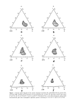
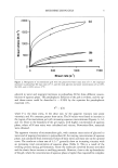
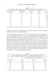
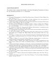
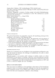
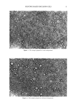
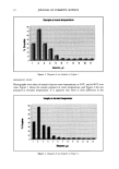
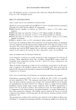
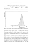
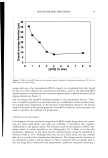
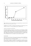
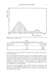
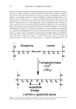
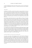
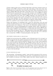
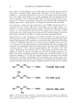
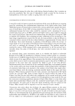
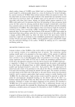
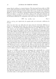
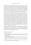
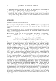
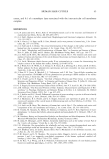
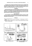
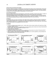
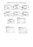
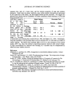
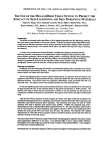
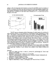
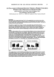
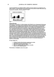
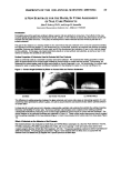
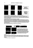
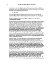
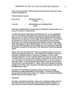
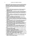
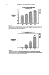
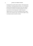
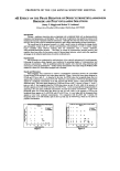
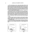
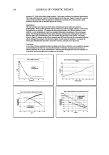
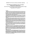
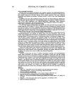
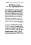
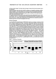
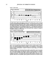
PREPRINTS OF THE 1998 ANNUAL SCIENTIFIC MEETING 51 To determine the types of interactions occurring between the polymer and clay, the absorbance bands for the edge (3620 cm ']) and face (1000-1200 cm ']) of the clay, the NI-I2-stretching region (3000-3750 cm -]) and the N-R3 + deformation band (1536 cm ']) of the polymers were monitored for peak shifts or the introduction of new peaks. From this analysis it was found that H-bonding and electrostatic interactions contribute to polymer adsorption primarily along the negative face surface of the clay. Conclusions H-bonding and cationic interactions of the polyacrylamide homo- and copolymers have been shown to contribute to the adsorption process. The PAIn acts as a H-bonding donator and adsorbs along the edge surface of the clay. At complete surface coverage changing the pH of this system alters the polymer conformation. In contrast, adsorption of PAmMaap Quat onto montmorillonite clay primarily occurs along the face surface of the clay through both H-bonding and electrostatic interactions while a rather flat or hindered conformation of the polymer is observed at each pH studied. Acknowledgements The authors gratefully acknowledge William Jarrett for his contribution in setting up the NMR experiments and Felicia Fye and Amy Marks for their work on generating the adsorption isotherms used for these studies. The authors would also like to thank Southern Clay Products for funding and supplying the montmorillonite clay used in these studies. References Bottero, J.Y. Bruant, M. Cases, J.M. Canet, D. and Fiessinger, F. J. Colloid Interface Sci., 124:2, 515, (1988). Webb, S.W. Stanley, D.A. and Scheiner, B.J. U.S. Dept. of the Interior, Bureau of Mines, Government Document 128.23:9036 (1986). Figure 1. ]3C NMR of PAm/Clay Complexes at Complete Surface Coverage 13340. 3200 INHz ntislob I NH, I I (2 Iqu•al©nt hazy#) I Figure 3. FTIR Spectra of PAm Figure 1. FTIR Spectra of Montmorillonite Clay claP/•dge: AI-Mg-OH o• • 4 4 • f•: S•O • O O, • 2. 2 • 115.189 139.1• 1•193 •Od• O.• 5.• •.• • Ibl blrld (cm) = pelk mlx,*-,- -• - pelk ftmx,-'--,- -• pH 3 pH 7 pH 10 Figure 4. Shifts in the PAm/Montmorillonite Clay Complex Spectra

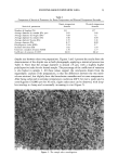
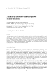
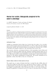
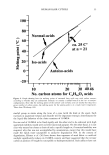
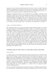
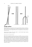
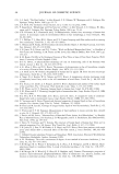
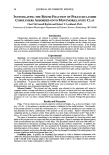
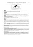
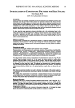
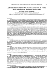
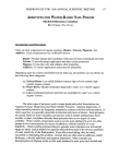
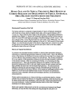
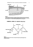
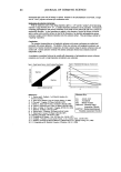
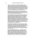
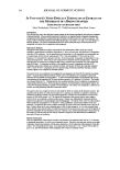
52 JOURNAL OF COSMETIC SCIENCE OIL EMULSIFICATION BY [}-CASEIN, POLY(L-ASPARTIC ACID), MANDUCA SEXTA APOLIPOPHORIN III, AND A SOYBEAN VACUOLAR PROTEIN Robert Y. Lochhead, Monica Tisack-Kathman i, Gordon Cannon and Charles L. McCormick University of Southern Mississippi, Department of Polymer Science, Hattiesburg, MS 39406 Introduction: Polymeric emulsifiers based upon hydrophobically-modified, cross-linked polyacrylic acid were introduced to the cosmetic industry a decade ago • 23 n. These emulsifiers have found utility in the delivery of emollients, barrier oils and sunscreens to the skin surface from emulsion formula- tions. The most stable emulsions are formed at polymer concentrations in excess of the critical overlap concentration and these emulsifiers appear to function by a mechanism in which the oil is trapped within hydrophobic •holcsf in a hydrophilic matrix 567 8 9. However, the design of synthetic polymeric emulsifiers is far from being understood, and many hydrophobically-modified hydrophilic polymers have been screened with little suc- cess as emulsifiers. It appears that a polymcric emulsifier must be capable of being adsorbed with sufficient rapidity at the oil/water interface. Moreover, the adsorbed polymer must be adsorbed in a preferred conformation and must interact throughout the continuous phase to confer a yield stress sufficient to prevent creaming of the emulsion. In Nature, proteins are commonly used to stabilize oil and fat droplets in the blood of animals. the lymph of insects and the sap of plants. The emulsion droplets found in plants are oil bodies. The proteins that stabilize these oil bodies are oleosins. In insects the stabilizing proteins are termed apolipophorins. This study was aimed at gaining an understanding of natural emulsification in order to guide the syntheses of future poly- meric emulsifiers. Soybean Oleosins: Plant oil bodies are termed oleosornes and the proteins which stabilize these oleosomes are termed oleosins. The oleosomes consist of three struc- tural constituents: triglycerides in the center, surrounded by a phospholipid layer and an external sheath of oleosins.(Figure I ). Figure 1. The structure of an oleosome Oleosins are alkaline proteins having molecular weights in the range 15,000 to 26,000 •ø. All oleosins possess three characteristic regions: a 40-60 amino acid N-terminal region, a 68-74 amino acid middle region which is hydrophobic, and a 33-40 amino acid C-terminus moiety. The N-termi- nal moiety is amphipathic. Its conformation is believed to be helical and it is suspected that this region interacts with the surface of the oil body. Tzen n determined that the central hydrophobic segment was arranged in an anti-parallel O-strand conformation which is suspected of penetrating the triglyceride core. This conformation is not certain •2 t3. Soy Vacuolar Protein A 34,000 g/mole protein, (Soy 34), is one of 4 major proteins found during the isolation of oil bodies from mature seeds an. This vacuolar protein is located in the storage vacuoles and immunocytochemical evidence indicates that Soy 34 associates with oil bodies only after disruption of seed cells •5. This protein consists of 257 amino acid residues and it does not possess the conserved hydrophobic portion which is typical of oleosins. Soy 34 does possess a periodic distrubution of hydrophobes. Avoliooohorins In insects, the proteins which stabilize the oil particles are known as Apolipophorins. Apolipophorin III is found in adult insects 'free' in the hemolympht6 •7. Insect Apolipophorin III was chosen for this study because of the apparent reversible association with hemolymph and lipophorins. The Apolipophorin III from Manduca Sexta (tobacco horn worm) has been studied fairly extensively, and, for that reason, this protein from this species was chosen for this study. Apolipophorin III is a 18,494g/mole water-soluble polypeptide which consists of 161 amino acid residues as. The protein conformation is believed to exist as a globular five-helix bundle. Each helix is amphipathic and the hydrophobic portions of each helix orient to the interior. ' This study con.ducted as partial requirement for the degree of Ph.D. Monica Tisack-Kathman, 1998, University of Southern Mississippi
Purchased for the exclusive use of nofirst nolast (unknown)
From: SCC Media Library & Resource Center (library.scconline.org)