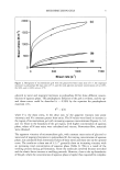
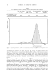
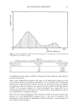
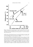
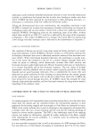
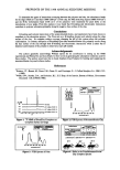
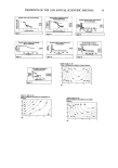
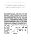
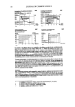
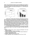
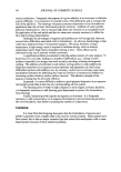
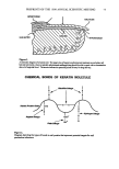
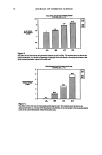
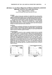
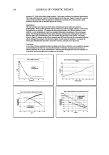
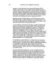
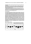
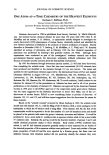
PREPRINTS OF THE 1998 ANNUAL SCIENTIFIC MEETING Ob•iective The goal of this research is to evaluate and gain insight into the structure-interfacial property relationships that exist in natural emulsifiers. The long term goal is to use this knowledge to design highly efficient smart polymeric emulsifiers. Experimental Materials Oleosins and vacuolar protein were isolated and purified by grinding soybean seeds in a buffer, centrifugation, and extraction of the supernatant with diethyl ether, followed by ethanol chloroform and further centrifugation to obtain an oleosin pellet. Isolation of the 34kD protein was achieved chromatographically using a DE52 urea column and varying salt concentration of the mobile phase. The 34kD protein was identified by SDS-PAGE. Apolipophorin III was obtained by recombitant synthesis in E. Coli lysogens •9. The cDNA library from the Manduca Sexta was a gift from Dr. Robert Ryan of the University of Alberta, Canada. Poly Na+ (L-Aspartic Acid), MW =15-50kD, and O-Casein, 90%, mw 23982 g/mole, were purchased from Sigma and used as received. Cis- Patinatic acid, MW 276g/mole, was purchased from Pierce and used as received. Methods Equilibrium and dynamic surface and interfacial tensions were measures using a Kruss TM k12 Tensiometer with Wilhelmy Plate and DeNouy Ring attachments. Emulsification experiments were conducted in accordance with regression analysis using an orthogonal factorial design TM. The independent vari- ables wet part and protein concentration. The dependent variable was moles of oil emulsified per mole of protein in solution. Emulsion experi- ments were conducted by placing 0.6ml of protein solution into a small vial and evaluating the molar quantity of otl which could be emulsified at saturation. Surface Hydrophobicio' of the proteins was determined using the surface fluorescence probe, cis-parinaric acid. Fluorescence intensity was mea- sured using an Edinburg FS900CDT T-geometry fiuorimeter. The excitation frequency was 325nm and emission was monitored at 420 nm. Initial slopes of fluorescence intensity versus protein concentration yielded the effective (or surface) hydrophobicity. Equilibrium Phase Diaeratns Emulsions of cyclohexane or tetradecane in water were prepared using the polymeric emulsifiers at various concentrations. The phase behavior was continually assessed over several months. Results and Discussion O-Casein was chosen as positive control. This protein is commonly used as an emulsifier in the food industry2L This protein exists as a disordered coil containing no disulfide bridges and approximately 10% •t-helices Lysozyme was chosen as a control. Lysozyme is an ellipsoidal molecule having dimensions 3nm x 3nm x 4.5nm. The protein contains many heli- cal segments and a three-stranded antiparallel O-sheet. The tertiary structure is locked in place by four disulfide bridges, Lysozyme is a rigid mole- cule in which the nonpolar groups are orientes to the interior of the molecule and the exterior is hydrophilic. The negative control was poly(aspartic acid) which is a highly water-soluble polypeptide. Surface Hvdroohobocities The fluorescence intensities of samples containing cis-parinaric acid are shown in figures 3 thro' 7 for aqueous solutions of the proteins studied. As expected, the lysozyme control showed no surface hydrophobicity, as did the poly(aspartic acid) control at pH's 8 and 12. It is interesting that poly(aspartic acid) showed significant surface hydrophobicity at pH 4, when it is relatively undissociated. B-casein displayed moderate hydropho- bicity. The 34kD Vacuolar Soy Protein displayed pronounced surface hydrophobicity at pH 12, but negligible surface hydrophobicity at pH Apolipophorin shows significant surface hydrophobicity at all pH's, with pH 12pH8~ pH4.

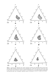
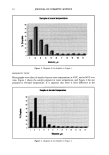
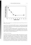
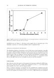
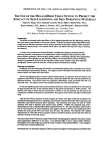
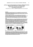
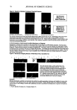
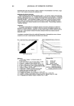
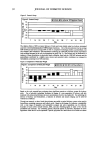
54 JOURNAL OF COSMETIC SCIENCE Interfacial Tensions at the Tetradecane/water interface The variation, with protein concentration, of interfacial tension at the tetradecane/water interface are shown in figures 8 through 12. [•-Casein, Soy 34kD vacuolar protein and Apolipophorin III all adsorbed at the oil/water interface in approximately the same concentration range. The equilibri- um surface tension at surface saturation is approximately the same for all three proteins, but Apolipophorin displays a relatively large dependence on solution pH, with acid and alkaline conditions favoring adsorption over neutral pH conditions. Rates ooeAdsorDtion at the Surface: [5-Casein, Soy 34kD Soy Vacuolar Protein and Apolipophorin III, all adsorb rapidly at the interface, lowering the surface tension by -20 mN/M. As expected, the rigid protein, lysozyme adsorbs and rearranges extremely slowly at the interface, taking weeks to reach equilibrium. Once adsorbed, Apolipophorin III does not desorb upon dilution as shown in figure 15. It is notable that the dilution was conducted sequentially over a period of one week and, therefore, it is reasonable to assume that the protein had ample time to re-equilibrate. This indicates that the inter- facial conformation and packing of this protein is probably thermodynamically favored over the solution conformation. Emulsification: The emulsification ability of the proteins are presented as contour diagrams (figures 16 through 18), which show the amount of oil emulsified as a function of protein concentration and pH. The emulsifiers can be ranked as the Apolipophorin being slightly better than the Soy 34kD protein which is slightly better than [5-Casein. It is interesting that [•-Casein emulsifies best at about pH 8.5, but the Soy 34kD Vacuolar Protein emulsifies best under alkaline conditions and the Apolipophorin III under acid conditions. Conclusions: 1. Lysozyme is a rigid protein having a hydrophilic exterior. It adsorbs weakly and slowly at aqueous interfaces and this protein does not display the capability to emulsify oil in water. 2. f/--Casein, Soy 34kD Vacuolar Protein and Apolipophorin III are all flexible amphipathic protein molecules that are readily and rapidly adsorbed at aqueous interfaces. These proteins are capable of emulsifying oil in water. 3. [•-Casein emulsifies best at about pH 8.5 4. Soy 34kD Vacuolar Protein emulsifies best at pH 12. 5. Apolipophorin III emulsifies best in acid conditions and, once adsorbed at the oil/water interface, the molecules of this protein reside at the interface even under conditions of high dilution with water or buffer solution. 6. Optimum emulsification is found for these protein emulsifiers under conditions that favor surface hydrophobicity, more rapid adsorption and more pronounced lowering of the oil/water interfacial tension. Surface Hydrophobiclty of B-C•sein 3 Protein concn, Surface Hydrophobicity of Lysozyme a= o --- , '- t• pill2 I S 10 15 Prokin coach, •/I Fi•ar• 4 Surface hydrophoblclty of 34kD Vicuoler Soy Protein 1500 o '":g ,, , , o 0.5 1 1 protein conch. 9/!- Figure 7
Purchased for the exclusive use of nofirst nolast (unknown)
From: SCC Media Library & Resource Center (library.scconline.org)