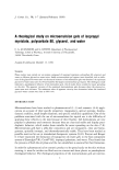
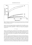
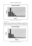
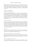
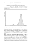
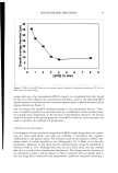
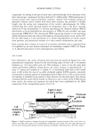
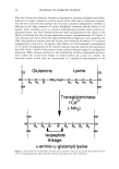
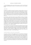
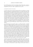
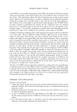
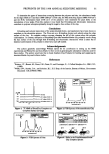
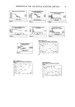
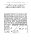
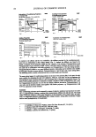
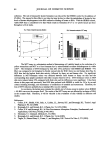
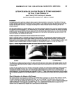
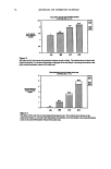
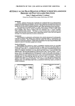
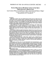
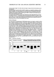
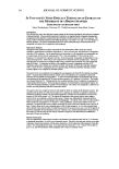
PREPRINTS OF THE 1998 ANNUAL SCIENTIFIC MEETING 63 A COMPARISON OF SKIN VIABILITY ASSAYS FOR IN VITRO SKIN ABSORPTION/METABOLISM STUDIES H.L. Hood • and R.L. Bronaugh z •Bristol-Myers Squibb, Clairol Division, Stamford, CT 2Office of Cosmetics and Colors, Food & Drug Administration, Washington, DC Introduction If metabolism is to be assessed during an in vitro percutaneous absorption study, it is essential that skin viability be maintained. It has been demonstrated that the viability of skin can be maintained for up to 24 hr in flow-through diffusion cells by utilizing a physiological receptor fluid with a carbohydrate energy source, such as HEPES-buffered Hanks' balanced salt solution (HHBSS) •. Water-insoluble compounds may not partition freely from excised skin into the aqueous HHBSS receptor fluid:. Therefore, to increase the aqueous solubility of lipophilic compounds in the receptor fluid 4% bovine serum albumin (BSA) is routinely added to HHBSS in our laboratory. A number of tests have been developed for assessing skin viability in vitro 3. Anaerobic glucose utilization (or the lactate assay) is the method employed most frequently in our laboratory as an indicator of tissue viability. Recently, however, we have observed that the addition of 4% BSA to HHBSS results in lower levels of measurable lactate in the receptor fluid. The objective of this study was to develop an alternate means of measuring skin viability that is unaffected by BSA. The MTr assay 4, which determines the presence of viable cells with metabolically active mitochondria, was evaluated as an alternative to the lactate assay. Viability of skin was measured with both the lactate assay and MTr assay following perfusion with HHBSS and HHBSS + 4% BSA. Species differences in the two assays were also examined using hairless guinea pig, fuzzy rat, and human skin. Methods Perfusion Study. Skin perfusion studies were performed in Bronaugh flow-through diffusion cells •. Dermatomed skin sections were placed in diffusion cells stratum comeurn-side up and skin surface temperature was maintained at 32 0 C. Receptor fluid was collected at a flow rate of 1.5 ml/hr in 6-hr fractions for 24 hr. Perfusion was ceased at various time intervals and skin viability was determined by the lactate assay or the MTr assay. Lactate Assay. Anaerobic glucose utilization in skin was determined by measuring lactate in the collected receptor fluid fractions with a Sigma © diagnostic kit. Briefly, a 0.20 ml aliquot from each 6-hr fraction was mixed with 2.8 ml of assay reagent. Samples were incubated in a water shaker bath at 37 øC for 45 min. The UV absorbance of the reaction mixture at 340 nm was measured spectrophotometrically and --'as proportional to the amount of lactate originally present. MTr Assay. MTr was dissolved in HHBSS at 2 mg/ml and warmed to 37 øC. At specified time intervals, the area of skin perfused was cut and placed into a 6-well tissue culture plate. To each well 2 ml of MTr solution was added and the plate incubated for 2 hr at 37 øC and 90% humidity on a rotating platform. Fresh control skin was dermatomed and also assayed after equilibrating in I-IHBSS for 20 min. After incubation the MTr solution was removed and each skin section was washed with phosphate-buffered saline. Isopropanol was added to extract the blue MTr-formazan crystals from skin by agitating plates on rotating platform at room temperature for 1 hr. Two ml of extract was diluted with 2 ml of isopropanol and the absorbance was measured at 540 nm. Results and Discussion The rates of anaerobic glucose utiliza, tion from perfusion of HGP skin with HHBSS and HHF• •' + ß 4% BSA are illustrated in Fig. 1 by plotting the micromoles oflactate produced per hour in each 6-hr

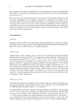
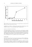
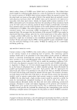
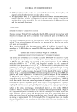
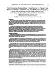
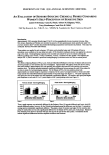
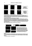
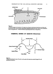
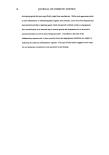
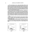
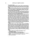
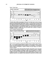
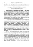
64 JOURNAL OF COSMETIC SCIENCE collection. The rate of measurable lactate formation was reduced for the HHBSS control by the addition of 4% BSA. The reason for this effect is not clear but may be due to either the metabolism of lactate by low levels of lactate dehydrogenase in the BSA utilized or binding of lactate to BSA. With the HHBSS control, viable HGP skin is considered to be that which results in formation of 0.7 to 1.0 [tmole lactate per hour throughout a 24-hr study. 1.0 6 12 18 34 •g z Ir•npa:• arabs • Mrr as? • zm The MTr assay is a colorimetric method of determining cell viability based on the reduction of a yellow tetrazolium salt MTr to a blue formazan dye by mitochondrial succinate dehydrogenase in viable cells n'6. The formation of MTr-formazan by skin after 24-hr perfusion with HHBSS and HHBSS + 4% BSA was compared to its formation by freshly excised skin for HGP, fuzzy rat, and human skin (Fig. 2). HGP skin had the highest fresh skin activity, followed by fuzzy rat and human skin. No significant difference in MTr-formazan values was obtained between fresh human or fuzzy rat skin and the corresponding skin samples perfused with either receptor fluid for 24 hr. MTr-formazan values in HGP skin were reduced about 25% compared with fresh skin and the differences were significant. It has been shown in this paper and previously • that HGP skin perfused with HHBSS is viable for 24 hr by several criteria including histology, and maintenance of metabolism of estradiol and testosterone. Therefore, the loss of MTr reduction probably has a minimal effect on skin viability. In conclusion, the lactate assay remains the viability test of choice except in studies where HHBSS + 4% BSA serves as the receptor fluid. The results of the MTr assay are not affected by inclusion of BSA in the receptor fluid. Therefore, it will be valuable in the evaluation of skin viability in diffusion cell studies. References: 1. Collier, S.W., Sheikh, N.M., Sakr, A., Lichtin, J.L., Stewart, R.F., and Bronaugh, R.L. Toxicol. Appl. Pharmacol. 99, 522 (1989) 2. Bronaugh, R.L. and Stewart, R.F.J. Pharm. Sci. 73, 1255 (1984) 3. Collier, S.W. and Bronaugh, R.L. In Vitro Percutaneous Absorption: Principles, Fundamentals, and Applications, CRC Press, Florida,31-49 (1991) 4. Mosmann• T. J. Immunol. Methods 65, 55 (1983) 5. Bronaugh, R.L. and Stewart, R.F.J. Pharm. Sci. 74, 64 (1985) 6. Triglia, D., Sherard Bma, S., Yonan, C., and Naughton, G.K. Toxicol. In Vitro 5, 573 (1991)
Purchased for the exclusive use of nofirst nolast (unknown)
From: SCC Media Library & Resource Center (library.scconline.org)