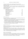
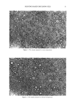
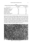
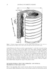
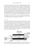
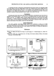
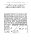
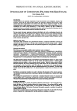
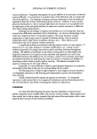
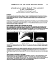
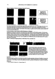
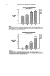
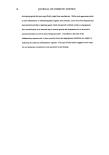
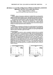
PREPRINTS OF THE 1998 ANNUAL SCIENTIFIC MEETING 61 THE USE OF THE MELANODERM TISSUE SYSTEM TO PREDICT THE EFFICACY OF SKIN LIGHTENING AND SKIN DARKENING MATERIALS Marie K. King,* M.S., Michael Caswell,* Ph.D., Hila A. Blochowitz,* M.S., Karen Adams,* M.S., James A. Greene, + M.S., and Richard L. Roberts, # Ph.D. *Stephens & Associates, Inc., Carrolton, Texas +Shaklee Corporation, Hayward, California #R.L. Roberts Product Development Consultants, Germantown, TN Introduction The ability to accurately predict the efficacy of skin lightening materials and skin darkening materials has, until recently, been limited to clinical test methods. With the introduction of the MelanoDerm tissue system from the MatTek Corporation and the protocol described in this paper, efficacy of these materials are effectively assayed using in vitro methods which offer a cost effective and rapid first step in screening test materials. A variety of raw materials and finished formulas, including skin lightening materials and skin darkening materials, were assayed in the MelanoDerm system to determine their potential to either prevent melanin production, remove melanin already present, or artificially stimulate tissue pigmentation. Measurement of skin lightening or skin darkening efficacy was accomplished by directly measuring changes in pigmentation of the tissue atter exposure to these various materials using three endpoints: photography, sodium hydroxide extraction of tissue melanin and Mexameter readings. Materhis and Methods The efficacy of the skin darkening test materials was evalunted by applying 100/zi of product to six tissues and incubnting for approximately 48 ho•rs. The negative control was treated with deionized water and the positive control was treated with 5mM DOPA. For the skin lightening assay to remove melanin already present, six tissues for each test material were exposed to 5mM DOPA for approximately 48 hours, rinsed, then dosed with 100/zl of test material for an additional 411 hours. The positive conuol was exposed to 5mM DOPA, rinsed, and dosed with 100/zl of deionized water. The negative control was exposed and dosed only with deionized water. For the skin lightening assay used to prevent melanin production, tissues in replicates of six were exposed for approximately 48 hours to test materials diluted to the desired concentration in DOPA solution. The dilutions were made to ensure that the concentration of DOPA in the test materials was the same as the concentration of DOPA in the positive control (SmM). The negative control for this procedure was deionized water. After the final 48 hour exposure for each procedure, the tissues were again rinsed and the melanin was quantitated photographically (all six replicates), instrumentally using the Mexameter (three replicates), and chemically using NaOH extraction (three replicates). The remaining three tissues not used for NaOH exlraction were used to determine post exposure tissue viability. Viability was measured using 3-(4,5- dimethylthiazol-2oyl)-2,5-diphenyltetrazolium bromide (MTT) (!=4). The mean absorbance of each well at $70 nm was divided by the mean absorbance of the negative controls at 570 nm and multiplied by 100 to calculate the mean percent MTT conversion value for each test material. Test materials with viability measurements of50% at•er 48 hours exposure would be considered cytotoxic and the data from these test materials would be viewed with caution. The Zeiss DermaVision TM Stereomicroscope/Vid• System with photographic capability was used to visually assess pigmentation diff, zrences in the tissue. Cross polarized light is used to reduce surfrice reflection. Photographs of the video images are printed for visual evaluation of test material efficacy. Color changes in the tissues were evaluated qualitatively. The Mexameter is an instrument designed to directly mensure the degree ofmelanogmesis and erythema seen

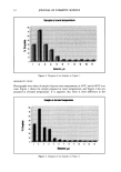
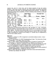
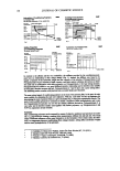
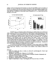
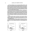
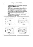
62 JOURNAL OF COSMETIC SCIENCE in viable tissue. The probe of the Mexameter emits the light of three defined wavelengths: 568nm, 660nm, and 880nm (two wavelengths are used for each endpoint). The receiver measures the light reflected by the tissue and, as the quantity of emitted light is defined, the quantity of light absorbed by the tissue can be calculated (5). This instrument is commonly used in clinical trials and has been adapted for use in the laboratory study to assess differences in melanin production compared to controls. Results obtained using the Mexameter from tissues exposed to test materials were compared to the negative and positive controls using a t-test (p0.05). The amount of melanin present in tissue can also be quantified by extracting with 2 N NaOH (6-7). Tissues were placed in 0.5 ml of 2 N NaOH and incubated for approximately 48 hours with mechanical agitation to exitact the melanin. Aliquots from each replicate were read spectrophotometrically at 410 nm. The mean absorbance of the six replicates was divided by the mean absorbance of the negative control and multiplied by 100 to calculate the mean percent for each test material. The mean percent value of each test material was then compared to the mean percent value of the positive control using the t-test (p0.05). Results The results and conclusion of this study will be presented and discussed at the Society of Cosmetic Chemists' December meeting. Refereuces !. Oslxrne, R. and M.A. Perkins, The Procter & Gamble Company. Evaluation of human skin cell cultures for in vitro skin irritancy testing. In VitroToxicology: Mechanisms and New Technology. New York: Mary Ann Liebert, Inc. 8:317-324. (1991). Carmichael, J., W.G. Degra• A.F. Gazdar, J.D. Minna, and J.B. Mitchell. Evaluation of a tetrazolium-based semiautomated colorimetric assay: assessment of chromosensitivity testing. Cancer Res. 47:936-942. (1987). Triglia, D., S.S. Braa, T. Donnelley, I. Kidd, and G.K. Haughton, Marrow Tech, Inc. A three dimensional human derreal model substate for in vitro toxicological studies. In Vitro Toxicology: Mechanisms and New Technology. New York: Mary Ann Liebert, Inc. 8:351-362. (1991). 4. MatTek product literature. 5. Courage+Khazaka (Mexameter) product literature. Talwar, H.S., C. Griffiths, G. Fisher, A. Russman, K. Krach, S. Benrazavi, and J. Voorhees. Differential Regulation of Tyrasinase Activity in Skin of White and Black Individuals In Vivo by Topical Retinoic Acivl J. Invest. Dermat. 100:800-802. (1993). Iozumi, K., G. Hoganson, R. Pennel!a, M. Everett, and B. Fuller. Role of Tyrosinase as the Determinant of Pigmentation in Cultured Human Melanocytes. J. Invest. Dermat. 100:806-808. (199S).
Purchased for the exclusive use of nofirst nolast (unknown)
From: SCC Media Library & Resource Center (library.scconline.org)