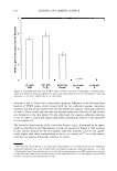
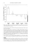
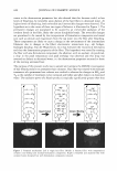
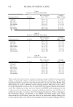
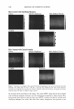
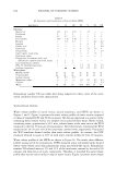
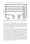
604 JOURNAL OF COSMETIC SCIENCE site was evaluated using various in vivo measurements (see the following three sections) at baseline (prior to treatment). Volunteers with TEWL values outside the normal range ( 10 g/m2hr) were excluded. Subsequently, the patch containing one of aqueous con tacting solutions i, ii, iv, or v was applied to the skin test site for five hours on Day 1. After approximately 18-20 hours on Day 2, the skin test site was re-evaluated. Some of the skin test sites were not exposed to the patches, and were left untreated and non-occluded to serve as the in vivo control (see above). The in vivo measurements were reported as deviations from the baseline measurements. Measurement of transepidermal water loss using an evaporimeter. Transepidermal water loss (TEWL) measurements provide a noninvasive instrumental assessment of the skin barrier function in vivo. Specifically, skin barrier perturbation may lead to a disruption of the intercellular lipid bilayers in the SC, thereby resulting in elevated water loss rates. Such elevated water loss rates can, in turn, lead to the skin becoming dry and chapped, thereby enhancing skin dryness (20-24). The TEWL measurements were made using an evaporimeter (CyberDERM, Inc., Broomall, PA) with probes manufactured by Cortex Technology (Hadsund, Denmark). This instrument is based on the vapor pressure gradient estimation method designed by Nilsson and initially utilized by the Servo Med Evaporimeter (25). The probes contain two sensors that measure the temperature and relative humidity at two fixed points along the axis normal to the skin surface. This arrangement is such that the device can electronically measure a value that corresponds to evaporative water loss from the skin surface expressed in g/m2hr. Additional details on the TEWL measurements using an evaporimeter can be found in references 26 and 27. The TEWL measurements were conducted following a 15-30-minute acclimation pe riod in a controlled environment, with the relative humidity maintained at less than 50% and the temperature maintained at 21 ° ± 1 °C. At baseline prior to treatment, TEWL measurements were conducted for each of the skin test sites. Any individuals .with TEWL values outside the normal range ( 10 g/m2hr) were excluded at this time. The test formulations were then applied to the test sites under occlusive soap chambers. On Day 2 (approximately 18-20 hours after patch removal), TEWL measurements were conducted for each of the skin test sites as described above. Evaluation of visual skin dryness by an expert grader. The visual skin dryness on the volar forearm test sites of each volunteer was evaluated by an expert grader using the grading scale described in Table I. Intermediate grades were allowed so that finer distinctions could be made. To conduct the study in an objective manner with no biases, the expert Table I Expert Grader Score System Used to Determine Visual Skin Dryness as Part of the In Vivo Soap Chamber Skin Barrier Measurements Grade 0 2 4 6 8 Description None Slight flaking/uplifting of flakes (patchy and/or powdered appearance) Moderate flaking/uplifting of flakes (uniform) and/or slight scaling Severe flaking/scaling, uplifting of scales and/or slight fissuring Severe scaling/uplifting of scales with severe fissuring/cracking

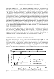
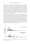
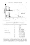
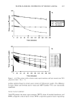
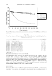
RANKING OF SURFACTANT-HUMECTANT SYSTEMS 605 grader was not made aware of the contents of the patches containing aqueous contacting solutions i, ii, iv, and v. One should note that the range, 0-2, included all the visual skin dryness scores reported by the expert grader (see Table I). Although these scores corre spond to the mild range, the expert grader was nevertheless able to effectively discrimi nate between the observed low levels of visual skin dryness. Measurement of skin erythema using a chromameter. Skin erythema was measured instrumen tally by a Minolta CR-200 chromameter that is based on a standardized reflectance technique using a tristimulus system (18). The tristimulus system makes use of color reading values on three independent axes: (i) L * axis, reflecting the tone of lightness/ darkness, with higher values indicating lightness and lower values indicating darkness (ii) a* red/ green axis, reflecting the extent of redness/ greenness, with higher values indicating more red tone and lower values indicating less red tone and (iii) b* blue/ yellow axis, reflecting the extent of blueness/yellowness, with positive values indicating a yellowish tinge and negative values indicating a bluish tinge. Specifically, the color reading values were translated into the L *a *b* coordinates whose spacing correlates closely with color changes perceived by the human eye. For the evaluation of skin erythema using a chromameter, only values along the a* red/green axis that can capture the extent of redness (erythema) of the skin were considered. Sets of three a* readings from each of the volar forearm test sites were taken at baseline, as well as on Day 2 (approximately 18-20 hours after patch removal), and the average a* value was calculated for each site. Increased a* values along the red/ green axis, relative to the baseline measurements, indicate that the patch containing the aqueous contacting solution has induced skin redness (erythema) (18). Additional details can be found in references 18 and 19. THEORETICAL DEVELOPMENT OF AN IN VITRO TEST TO RANK AQUEOUS SURFACTANT-HUMECTANT SYSTEMS The central objective for developing an in vitro ranking metric is to rank aqueous surfactant-humectant contacting solutions i, ii, iv, and v, relative to the in vitro PBS control (iii), based on the extent of their perturbation to the skin aqueous pores. For this purpose, we chose skin electrical current induced by aqueous contacting solutions i-v, relative to the in vitro PBS control (contacting solution iii), as the preferred in vitro ranking test. Specifically, we adopted the following in vitro ranking metric (RM): IE RM= Ic (1) where E denotes the enhancer (that is, aqueous contacting solutions i, ii, iv, and v), C denotes the control (that is, the in vitro PBS control (iii)), IE denotes the skin electrical current induced by EJ and l e denotes the skin electrical current induced by C. Note that RM in equation 1 corresponds to the enhancement in the skin electrical current. The in vitro skin electrical current measurements ( 6,41) show that these measurements are: (a) extremely sensitive to small changes in the extent of skin barrier perturbation, (b) highly reproducible, and (c) simpler to implement, less time-consuming, and safer than typical skin permeability measurements, which make use of radioactive materials
Purchased for the exclusive use of nofirst nolast (unknown)
From: SCC Media Library & Resource Center (library.scconline.org)