127 PHYSALIS ANGULATE CREAM FOR SOLAR MELANOSIS IPL is high-intensity, polychromatic, noncoherent, and uncollimated. The treatment was idealized by Goldman in 1963 and improved utilizing the principles of selective photothermolysis. In IPL, selective destruction of a target pigment, called chromophore, is achieved by a specific light length with a minimal thermal effect at a distance (6–8). IPL is used in folk medicine as an antirheumatic, hepatoprotective, and in cases of jaundice (9), as a diuretic, trypanocide, cough suppressant, analgesic, and anti-inflammatory substance (10,11). Plants of the Solanaceae family are rich in polyoxygenated compounds that produce intact or modified structures of ergostane. These compounds are called vitasteroids (12). In P. angulata, phytosteroids are the most important compounds and are responsible for the anti- inflammatory and immunomodulatory effects (12). These phytosteroids have not yet been fully identified, although it is known that they are present in larger quantities in the leaves and the stem of the plant. P. angulata steroids such as vamolideo and fisangulideo were isolated from the leaves. Other phytosteroids and B, D, E, F, G, H, I, J, and K physalins were isolated from the stem. The fisangolide together with the epoxide D show anti-inflammatory activity similar to hydrocortisone in inflammatory processes (12). The extract of fruits and roots of P. angulata were found to have antimicrobial activity without any phototoxic effects (13). The aqueous extract of the P. angulata root shows efficacy in pain relief (14). Physalin E showed efficacy in improving dermatitis induced in animals (15) as well as the ability to inhibit myeloperoxidases, neutrophil migration, and the production of free radicals (16,17). In addition to phytosteroids, phytochemical studies reported the existence of other compounds such as alkaloids (18) and flavonoids (19) however, these results have not been fully identified. Moreover, P. angulata has also demonstrated antiseptic properties (20). The hydroalcoholic extract of P. angulata was shown to effectively modulate the production of inflammatory response mediators such as histamines, prostaglandin E2, interleukin-1 alpha, interleukin-6, and interleukin-10 in vitro (21). An experimental study of a cream containing the supercritical extract of P. angulata, in a culture of human keratinocytes exposed to sodium dodecyl sulfate (irritant surfactant), showed an ability to inhibit interleukin-1 alpha similar to that of dexamethasone 17-valerate (22). Considering the need to discover new anti-inflammatory drugs in dermatology and to explore the properties of P. angulata described in literature, the aim of this study was to evaluate the anti-inflammatory action of the cream containing 0.5% supercritical extract of P. angulata L. in comparison to 1% hydrocortisone. We observed this effect in an inflammatory process that occurs after IPL treatment of hand melanosis. MATERIALS AND METHOD EXPERIMENTAL PROCEDURE This was a double-blind, placebo-controlled clinical study conducted with 60 participants (n = 60) in a private dermatological office from July 01, 2019 to August 01, 2019 after approval by the Ethics Committee (CAAE 11089219.20000.0109). The study was conducted with participants who wished to undergo IPL treatment for cosmetic improvement of solar hand melanosis and consequently developed inflammation in both hands immediately after IPL application. The following inclusion criteria were applied: both genders were included, aged 26–73 years, in phototype II to IV, and all participants wished to be treated for solar melanosis

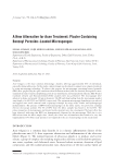
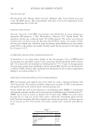
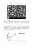
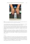
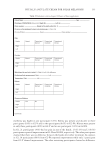
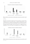
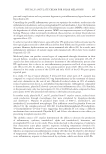
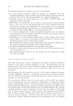
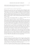
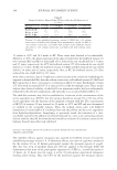
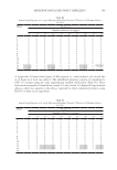
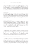
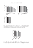
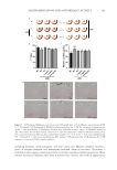
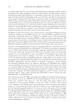
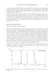
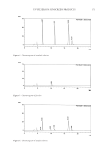
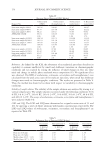
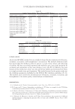
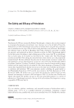
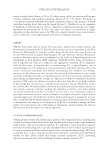
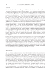
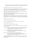
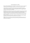
128 JOURNAL OF COSMETIC SCIENCE with IPL for better aesthetic results. The exclusion criteria included: participants who were smokers, diabetic, presented precancerous lesions during dermatoscopy, and had large discrepancy of lesions between the hands. Dermatoscopy was performed in all participants, with a Dermlite 3 Gen® portable dermatoscope with diode light and 10-times magnification, to rule out possible associations with precancerous lesions. Melanosis was marked on both the right and left hands of the 60 participants, with macules as similar as possible in color and diameter on both hands. This marking was performed to serve as a parameter for the analysis of erythema and temperature when the participants return after 48 h. This melanosis was properly identified in the photographs to facilitate findings at the time of reassessment Figures 1 and 2. Figure 1. G1 participant. (A) photo before treatment, (B) photo immediately after with the marking of the melanosis of both hands in which the measurement of perilesional erythema in mm was performed with the dermatoscope and where the temperature was also measured, and (C) 48 h after treatment where higher erythema can be seen on the left hand. Figure 2. G2 participant. (A) photo before treatment, (B) photo immediately after with the marking of the melanosis of both hands in which the measurement of the erythematous halo in mm was performed with the dermatoscope and where the temperature was also measured, and (C) – photo 48 h after treatment where a more intense inflammatory process is observed in the right hand with edema and blisters.
Purchased for the exclusive use of nofirst nolast (unknown)
From: SCC Media Library & Resource Center (library.scconline.org)