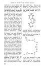
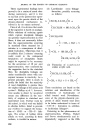
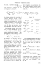
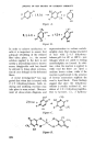
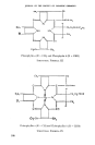
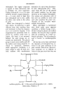
JOURNAL OF THE SOCIETY OF COSMETIC CHEIlilS'rS cluded in the case of cellulose and silk that the chain molecules con- stituting the fibre crystallites are as fully extended as possible •ø. This view was in agreement with the fact that on stretching fibres (1) the X- ray diffraction patterns remain un- changed (2) fibres do not recover their original length completely when the stretching force is re- moved. Stretching of cellulose and silk was, therefore, considered to occur by internal slipping of chain bundles over one another. Wool, on the other hand, does not behave in this way. Wool fibres in water ex- hibit long range elasticity, with the property of recovering their original length when the stretching force is removed. Furthermore, X-radio- grams of normal wool (a-keratin) are different from those of natural silk. However, as wool or hair is stretched, the normal X-ray photo- •aph gradually disappears and is replaced by a new one (/9-keratin) which is similar to that given by natural silk. It was concluded from this that stretched wool is built from fully extended polypeptide chains. When wool fibres which have been stretched in water at ordinary tem- peratures are released in water, they quickly recover their original length, and, at the same time, the normal X-ray pattern returns. This process can be repeated many times, show- ing that the transformations are re- versible. Since stretched wool was considered to consist of fully ex- tended protein chains, it was con- cluded that the unstretched fibre is built of the same chains in a regu- larly folded state. This was the position in the early 1930's, since which time much discussion has centred round the nature of the fold. The original hexagon fold of keratin: -- was later shown to be incapable of providing adequate space for accom- modating the amino acid residues, and the model was accordingly re- vised as show below•a: I co x.H H C--NH--CO--C R Rx NH I co RCH NH I co H Hx x C---CO--NH--C NH R R I It has been suggested by other workers, however, that a- and /•- keratin consist of many different types of fold, and it is believed that 162

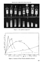
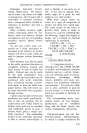
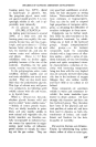
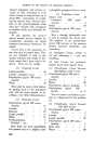
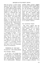
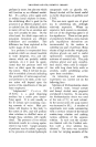
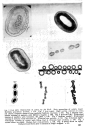
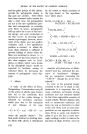
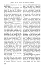
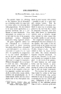
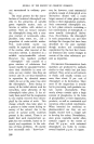
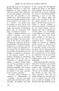
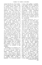
..::::'"'Q7 0 col o Fig. 1 (top leit): Destruction o[ cortex by 5% Na_oS. Note separation o[ cuticle layers II{olinsky guard-hair section). x 350. Fig. 2 (top right): High cystinc content o,f cortex md almost complete absence from medulla (Sullivan reaction). x 700. Fig. 3 (middle le[t): tntense staining of medulla with Millon's reagent. x 700. Fig 4 (bottom felt)' Preferent :taining of medulla with Safranine TS. pH7. x 150. Fig 5 (bottom centre): Preferent ,taining of medulla with Safranine B. (Russian hare section). x 150. Fig. 6 (bottom right): •referential staining of cortex with Kiton Red G (Russian hare section). x 150. Fig. 7 second down on right): Human hair cross-sections. x 150. Fig. 8 (third do•n on right): 12educed wool fibres after 5 minutes in Feigl's reagent. x 43. 163
Purchased for the exclusive use of nofirst nolast (unknown)
From: SCC Media Library & Resource Center (library.scconline.org)