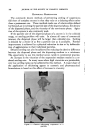
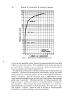
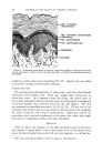
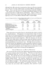
MUCOPOLYSACCHARII2ES IN HUMAN EPIDERMIS 155 this material was believed to have originated from the epidermal cells. This belief was based on the following finding: whole skin (epidermis -+- dermis) yielded considerably higher amounts of this mucin-like material than skin denuded of its epidermis. In the ensuing years more and more evidence began to accumulate indicating that in some epidermal structures and locations and under cer- tain conditions, mucopolysaccharides appeared to be consistently present. The evidence was based on histochemical observations and on direct chem- ical analyses. It is beyond the scope of this paper to discuss the specialized and often rather involved techniques which are used for the histochemical identifica- tion of mucopolysaccharides. For our purposes it will be sufficient to state that none of the histochemical methods are in themselves specific for mucopolysaccharides. However, when certain technical details are fol- lowed and when the effects of certain enzymes on the suspected material are observed, it is possible to conclude with a high degree of probability that the substance in question is indeed a mucopolysaccharide. One of the most characteristic staining features of mucopolysaccharides is their metachromasia, i.e. their ability to stain in a color different from the dye solution itself. For details the reader is referred to reviews in the field (3,4,5). Among epidermal structures, metachromatic material was found by Swedish scientists in some growing structures, such as the nail matrix (6), in the root sheaths of the hair, in epidermal hyperplasia, in precancerous and cancerous lesions (7, 8). More recently Montagna et al. published beautiful pictures, showing metachromasia in the external sheath of the hair follicle. Of great interest is also the intense metachromasia of the papillae of growing hair follicles (9). With the use of metachromasia and of other histochemical techniques, mucopolysaccharides were shown in the intercellular spaces of normal (9a) and pathologic epidermis (10) and in psoriatic epidermis (11, 12) and horny layer (13). CHEMICAL ANALYSES Hydrolysis of epidermal structures and extracts sets free the component elements of mucopolysaccharides. Building stones, such as hexosamine, hexose and uronic acid can be qualitatively and quantitatively analyzed. A mucoprotein was isolated from extracts of psoriatic scales by Roe (12) a similar substance could be extracted from scales o,f a patient'with ex- foliative dermatitis as well (14). Hexosamine, protein-bound hexose and glucuronic acid were found in homogenates of normal epidermis in higher concentrations than in blood serum (10) these compounds also occurred in hydrolyzed scales and scale extracts of patients with psoriasis (1,1).

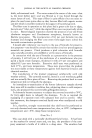
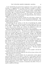
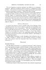
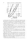
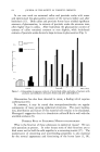
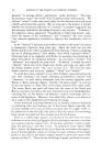
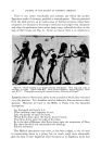
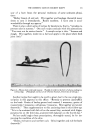
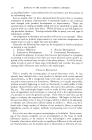
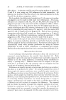
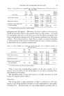
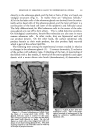
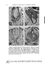
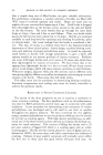
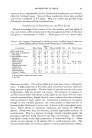
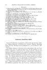
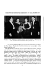
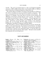
156 JOURNAL OF THE SOCIETY OF COSMETIC CHEMISTS In our own work we extracted callus and psoriatic scales with water and determined the glucosamine content of the extracts before and after hydrolysis (15). Both callus and psoriatic horny layer yielded significant amounts of glucosamine in extracts of psoriatic scales the amounts were often higher than in callus. After hydrolysis the glucosamine content of extracts of callus remained constant or rose slightly, while hydrolyzed extracts ofpsoriatic scales showed a large increase in glucosamine (Fig. 1). 120_ 100_ 0- Non-hydrolyzed extract I Hydrolsrzed extract i 2 3 4 5 6 CALLUS PSORIASlS EXFOL. DERM. Figure 1.--Glucosamine in aqueous extracts of pulverized callus and scales of patients with psoriasis and exfoliative derrnatitis before and after acid hydrolysis. Glucosamine has also been detected in sweat, a finding which requires confirmation (16). In summary, it may be stated that mucopolysaccharides are regular components of many growing epidermal structures. The exact localiza- tion, distribution and nature of this material still remains to be elucidated. Montagna's suggestion that it is chondroitin sulfate B fits in well with the available evidence (9). POSSIBLE ROLE OF EPIDERMAL MUCOPOLYSACCHARIDES What is the function of these substances in epidermal tissues? We can only speculate at present. In other tissues some of their functions are to bind water and to hold the cells together in a cementing matrix (17). The maintenance of cementing and waterbinding properties is also essential for the normal appearance and functioning of the horny layer (1, 18).
Purchased for the exclusive use of nofirst nolast (unknown)
From: SCC Media Library & Resource Center (library.scconline.org)