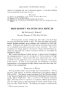
192 JOURNAL OF THE SOCIETY OF COSMETIC CHEMISTS (11) Herrmann, F., Prose, P. H., and Sulzberger, M. B.,Ibid., 21,397 (1953) (12) Harber, L. C., Herrmann, F., Mandol, L., and Sulzberger, M. B., Ibid., •9, 55 (1957). (13) Herrmann, F., Harber, L. C., Scher, R., Mandol, L.,/1. M./1./lrch. Derrnatol., 76, 282 (1957). (14) Wolf, J., Z. rnikroskop. anat. Forsch., 47, 351 (1940). (15) Szakall, A., Fette u. Seifen, 53, 399 (1951). (16) Pinkus, H., 2 t. Invest. Derrnatol., 16, 383 (1951). (17) Buckup, H., and Szakall, A., Berufsderrnatosen, 4, 1 (1956). (18) Herrmann, F., .drch. DerrnatoL SyphiloL, 200, 3 (1955). (19) Nicolaides, N., and Wells, G. C., •7. Invest. Derrnatol., 29, 423 (1957). (20) Montagna, W., "The Structure and Function of the Skin," New York, Academic Press, Inc. (1956). (21) Steigleder, G. K., and L6fFler, H., ,4rch. klin. u. exptl. DerrnatoL, 203, 41 (1956). (22) Steigleder, G. K., •7. Invest. Derrnatol., $1,29 (1958). (23) Kooyman, D. J., ./1. M. ./1. .drch. Derrnatol. SyphiloL, 25, 444 (1932). (24) Rothman, S., "Physiology and Biochemistry of the Skin," Chicago, University of Chi- cago Press (1954). RESPONSE OF THE HUMAN SEBACEOUS GLAND TO EXPERIMENTAL STRESS By JOHN S. STRAUSS, M.D.* Presented October 8, 1958, Seminar, New York City THE PURPOSE of this report is to detail the specific pathologic re- action patterns of the sebaceous glands following various experimental stresses. Furthermore, the responses will be correlated into a concise concept of response of the sebaceous gland after injury. However, before this is done, a few facts about the embryology, structure and function of the sebaceous glands must be reviewed. In embryonic life localized increased mitotic activity of the basal cell layer results in a bulging of the epidermal cells into the primitive derreal tissue. This is the primary epithelial germ which then grows down as a solid cord of basal cells. Part of the cord differentiates into a hair, other parts of this epithelial column form the sebaceous glands. Thus in embryonic life the entire pilosebaceous apparatus, including the hair and sebaceous gland arise directly from the epidermis. While the sebaceous glands vary greatly in size from area to area, the glands of the face and scalp are among the largest found in the body. Those in the scalp are associated with large terminal hairs, but on the glabrous portions of the face, particularly the cheek, the sebaceous glands reach enormous proportions and are the dominant structures in the follicles. Here the pilosebaceous duct leads * Dept. of Dermatology, Boston University School of Medicine--Massachusetts Memorial Hospitals, Boston 18, Mass. This study was supported under Grant E-1936C1 from the National Institutes of Health, United States Public Health Service.

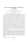
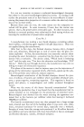
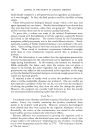
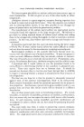
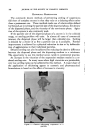
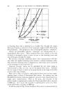
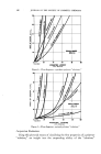
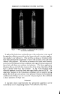
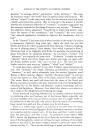
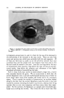
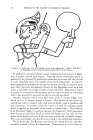
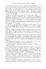
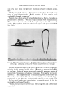
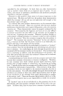
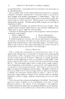
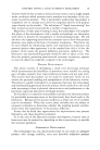
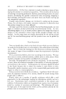
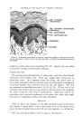
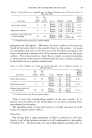
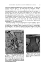
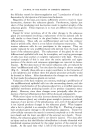
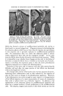
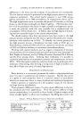
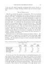
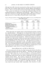
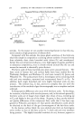
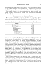
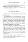
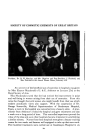
RESPONSE OF SEBACEOUS GLAND TO EXPERIMENTAL STRESS 193 directly to the sebaceous gland, and the hair or hairs, if they are found, are vestigial structures (Fig. 1). In reality these are "sebaceous follicles." All of the fat-laden cells of the sebaceous glands are derived from the mitot- ically active basal cells of the sebaceous gland, and the later cell layer is a continuation of the basal cell layer of the epidermis and follicular canal. The fully differentiated, fat filled sebaceous cells in the center of the seba- ceous glands are cast off to form sebum. This is called holocrine secretion. On histological examination, keratin-like trabeculae are also seen in many mature sebaceous cells. In other words, they are bipotential and still can produce keratin. On the other hand, the surface epidermal cells produce keratin as their main product, but also produce lipid material which is part of the surface lipid film. The following were among the experimental stresses studied in relation to changes in the sebaceous glands (1). 1) contact dermatitis, 2) occlusion of the surface with adhesive tape, 3) plucking of the hair, 4) puncturing of the follicle with a fine needle, 5) removal of the entire epidermis and upper dermis with a motor driven wire brush (dermabrasion), 6) destruction of Figure 1.--Normal pilosebaceous follicle from the cheek of a young adult male (X34). The dilated follicular canal is normal for this area. The sebaceous glands are very large and the follic- ular canal leads directly to them. On the left there is a small veilus-type telogen hair with evi- dence of new hair forming beneath this. The dark staining material in the follicular canal is made up of clumps of propionibacteria. ß .':.• .. .,,)' ::•.. • .•:.:.-..% %:.: ß zg• •--,• • •.. .. 3'- •.•.• - .:.. . .•,•.•...'•..•.• ,•.• :•' ? •: .. "% .... .. 3•: •"• '?'• .• ..... ... ', Figure 2.•Cheek 9 days after der- mabrasion (X52). This is the proto- typical example of replacement of the gland by primitive undifferentiated epithelial cells.
Purchased for the exclusive use of nofirst nolast (unknown)
From: SCC Media Library & Resource Center (library.scconline.org)