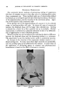
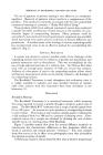
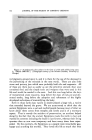
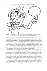
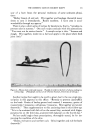
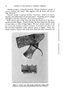
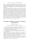
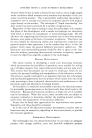
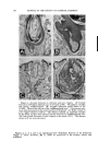
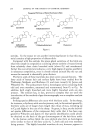
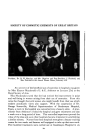
194 JOURNAL OF THE SOCIETY OF COSMETIC CHEMISTS the follicular mouth by electrocoagulation and 7) production of local in- flammation by the injection of irritants into the dermis. Regardless of the type, any injury, sufficiently severe to result in tissue destruction, destroys the sebaceous glands. Furthermore, injuries just short of that producing total destruction result in marked atrophy of the sebaceous gland. These responses in no way are different than that seen in any tissue. Except for minor variations, all of the other changes in the sebaceous gland are stereotyped involving a replacement of the fat-ladened cells by cells similar to those found in the gland before it shows any sebaceous differentiation. These cells are undifferentiated and look like ordinary basal or prickle cells there is a true failure of sebaceous maturation. The mature sebaceous cells do not participate in the response. They are merely replaced by new undifferentiated cells derived from the basal cell layer of the sebaceous gland. The replacement of the gland by the un- differentiated epithelial cells may be incomplete, and varying percentages of cells may show partial or complete replacement with lipid. The pro- totypical example of this is seen after the entire epidermis and upper portions of the dermis and cutaneous appendages are removed in derma- brasion. By five days many of the mature sebaceous cells are replaced by these "undifferentiated cells," by nine days the change is almost complete (Fig. 2). Actually these cells are a major contributor to the formation of a new epidermis and without them this plastic procedure probably would be doomed to failure. After dermabrasion the changes are reversible and by four weeks the sebaceous glands are normal. Variations of the basic response are common, resulting in proliferation of the undifferentiated epithelial cells into the surrounding dermis, and also complete transformation of the sebaceous gland into a stratified squamous epithelial membrane producing keratin as its product (squamous meta- plasia). However, since these changes occur principally after the pro- duction of dermal inflammation they will be discussed subsequently. While minor injury to the skin (contact dermatitis) produced no changes in the sebaceous gland, any stimulus great enough to promote a profound response of any portion of the pilosebaceous unit, even if the gland itself is not injured produces changes in the sebaceous gland. What happens after a hair is plucked is an example of this (Fig. 3). Plucking of anagen hairs produces great damage to the pilosebaceous apparatus. Except for a few cellular remnants the matrix is removed completely as is the internal root sheath and the lower one-third of the external root sheath. Never- theless, the sebaceous gland itself is not directly injured. Five days after an injury such as this the follicular infundibulum is already distended with keratinized squamae produced by the upper portion of the external root sheath. At the same time what is left of the lower portion of the hair

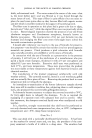
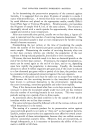
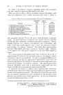
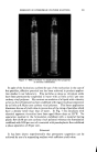
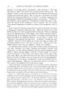
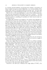
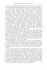
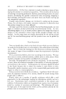
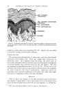
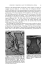
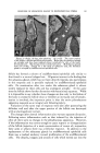
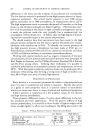
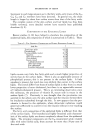
RESPONSE OF SEBACEOUS GLAND TO EXPERIMENTAL STRESS 195 .. . ... ..:&..• .. :•, .......... ,. , ...:• . :. -. t ::: ..,..:'"::'. .... ½..' - .? .... •':.'½. - •4 .: -' . -', - •,"½:' ::' .'.'. ' ..... "'.,f" .• ':% .':•':': : •'. :-'- '-T-' ." .•"½ :" t ': - ....... ? • •'"-' - : ½'-..':: :,.½•'1 .:.:... • •*.• -: ß • , . • ... • . . .., •..- . • -..f• •-: •:' -z• ß .'-• •:: •'.•'• -" • --• 'i': •" i -:% •'" :• •-"- ..... ¾ . • .....: ..: . ..•, •% .. • -•..,.•, ,.. ..... • • • • ß ... ...... . •. :. ,•: •. . .... : •. •.• .... •,: • • . .... •. * • ....... • • - • .•.. . • - • • • .... . ..... •. , -• •- '• -• • ß .-•, - ½.. . : - . . .•-•:.... ..:.. .... .. . •:.•.'• .... •.: ...•. •:- .½ • - ..• . --•:: .•.': 'c•- ..• *:' .... '- '-. •.•.:, --"::.:•S• • .... ,• • . •.• ..... • ..• •:.• .... • .... ½. -..._ . .• : -.... ß • .... • - .::.- •. ½ -.-'." •. •. • ß . • - . • . •.. Figure 3.--Scalp, 18 days after plucking. (g) (X33). The upper portion of the follicle is dilated and filled with keratin. Below this the sebaceous glands are atrophic and have been replaced almost completely by cells of the undif- ferendated type. The changes mimic comedo formation. (B) Sebaceous glands from 1A (X103). Except for a few nests of sebaceous cells, the shrunken gland is occupied by undifferentiated epithelial cells. follicle has formed a column of undifferentiated epithelial cells similar to that found in a normal telogen hair. Of greater interest is the finding that the sebaceous gland, which has not been directly injured, also participates in this response and is partially replaced by undifferentiated epithelial cells. On examination after two weeks the sebaceous gland is almost totally replaced by these cells and has undergone atrophy. At the same time the follicle shows further distention with keratinized squamae. While it is impossible to say whether these changes are due only to the failure of the hairs to sweep the follicle clean or whether an increased rate of keratin- ization is involved, the important point is that the entire pilosebaceous apparatus responds as an integral unit following injury. Variations of the same type of response were seen after puncturing the follicular wall and after the upper portion of the follicle was destroyed with an electrogalvanic current. The changes after derreal inflammation also warrant separate discussion. Following minor inflammation such as that induced by the injection of olive oil there were no changes in the pilosebaceous apparatus. However, if the inflammation was severe enough to cause rupture or disorganization of the follicle (injection of a weak concentration of croton oil, turpentine, fatty acids or sebum) there was a follicular response. In addition to the replacement of the sebaceous glands by undifferentiated epithelial cells there was a marked tendency toward proliferation of the undifferentiated cells. The sheaths, tongues and strands of cells which extend out into the
Purchased for the exclusive use of nofirst nolast (unknown)
From: SCC Media Library & Resource Center (library.scconline.org)