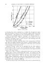
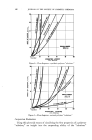
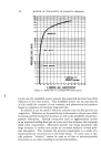
STRIPPED AND UNSTRIPPED HUMAN SKIN 191 rier /eve/ than for the intact surface. In 12 series of assays performed in seven psoriatics, the ratio of the medians for the stripped to the medians for the unstripped areas was greater than one and a half. There was no difference in the findings obtained in sites with psoriatic plaques (seven series) and those obtained in grossly unaffected skin areas (five series) of these patients. A median-ratio greater than one for "stripped to un- stripped" was observed also in a subject suffering from a skin ailment other than psoriasis, namely from congenital ichthyosiform erythroderma both ailments, however, have extensive parakeratosis in common. Another subject, suffering from extensive hyperkeratosis caused by a nevoid condi- tion, did not show a comparable ratio above one. As to the question of factors possibly underlying this reversal of the ratio of the acid numbers for barrier and intact surface, we might allude to the alternate possibilities of the existence of a microbial peculiarity on the one hand, an assumption favored by Rothman and his group, and of an intrinsic metabolic disorder on the other. We are inclined to speculate in the latter direction--in particular to attribute importance to several findings adding up to the hypothesis that there may be increased utiliza- tion of fatty acids for sterol synthesis in the outer horny layers. Summary 1) The quantity of ether-soluble substances on the skin ("casual level"), the acid number and the spreading index of these lipids were assayed in samples obtained from the intact skin surface and in samples obtained after stripping at the barrier level of the stratum comeurn. 2) The lipid quantity of the barrier level normally amounts to ap- proximately two-thirds to three-quarters of the quantity collected from the intact skin surface. 3) The acid number for the barrier level ranges by about 25 per cent below the acid number for the unstripped surface. 4) There is no difference in the spreading index for the two levels. 5) An acid number higher for the barrier zone than for the unstripped skin surface was found in the psoriatics thus far examined. REFERENCES (1) Herrmann, F., Coon, William, Harber, L., Scher, R. L., and Mandol, L., y. Soc. Cosmetic Chemists, 10, 88 (1959). (2) Szakall, A., Atrch. klin. exptl. Dermatol., 201,331 (1955). (3) Stupel, H., and Szakall, A., "Die Wirkung yon Waschmitteln auf die Haut," Heidelberg, Dr. Alfred H•thig Verlag (1957), p. 40. (4) Szakall, A., Berufsdermatosen, 6, 93 (1958). (5) Blank, I. H., y. Invest. Dermatol., 18, 433 (1952). (6) Blank, I. H.,Ibid., 21,259 (1953). (7) Blank, I. H., and Shappirio, E. B., Ibid., 25, 391 (1955). (8) Grueneberg, T., and Szakall, A., Atrch. klin. exptL DermatoL, 201,361 (1955). (9) Herrmann, F., and Prose, P. H., y. Invest. DermatoL, 16, 217 (1951). (10) Prose, P. H., Baer, R. L., and Herrmann, F., Ibid., 19, 227 (1952).

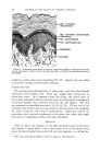
192 JOURNAL OF THE SOCIETY OF COSMETIC CHEMISTS (11) Herrmann, F., Prose, P. H., and Sulzberger, M. B.,Ibid., 21,397 (1953) (12) Harber, L. C., Herrmann, F., Mandol, L., and Sulzberger, M. B., Ibid., •9, 55 (1957). (13) Herrmann, F., Harber, L. C., Scher, R., Mandol, L.,/1. M./1./lrch. Derrnatol., 76, 282 (1957). (14) Wolf, J., Z. rnikroskop. anat. Forsch., 47, 351 (1940). (15) Szakall, A., Fette u. Seifen, 53, 399 (1951). (16) Pinkus, H., 2 t. Invest. Derrnatol., 16, 383 (1951). (17) Buckup, H., and Szakall, A., Berufsderrnatosen, 4, 1 (1956). (18) Herrmann, F., .drch. DerrnatoL SyphiloL, 200, 3 (1955). (19) Nicolaides, N., and Wells, G. C., •7. Invest. Derrnatol., 29, 423 (1957). (20) Montagna, W., "The Structure and Function of the Skin," New York, Academic Press, Inc. (1956). (21) Steigleder, G. K., and L6fFler, H., ,4rch. klin. u. exptl. DerrnatoL, 203, 41 (1956). (22) Steigleder, G. K., •7. Invest. Derrnatol., $1,29 (1958). (23) Kooyman, D. J., ./1. M. ./1. .drch. Derrnatol. SyphiloL, 25, 444 (1932). (24) Rothman, S., "Physiology and Biochemistry of the Skin," Chicago, University of Chi- cago Press (1954). RESPONSE OF THE HUMAN SEBACEOUS GLAND TO EXPERIMENTAL STRESS By JOHN S. STRAUSS, M.D.* Presented October 8, 1958, Seminar, New York City THE PURPOSE of this report is to detail the specific pathologic re- action patterns of the sebaceous glands following various experimental stresses. Furthermore, the responses will be correlated into a concise concept of response of the sebaceous gland after injury. However, before this is done, a few facts about the embryology, structure and function of the sebaceous glands must be reviewed. In embryonic life localized increased mitotic activity of the basal cell layer results in a bulging of the epidermal cells into the primitive derreal tissue. This is the primary epithelial germ which then grows down as a solid cord of basal cells. Part of the cord differentiates into a hair, other parts of this epithelial column form the sebaceous glands. Thus in embryonic life the entire pilosebaceous apparatus, including the hair and sebaceous gland arise directly from the epidermis. While the sebaceous glands vary greatly in size from area to area, the glands of the face and scalp are among the largest found in the body. Those in the scalp are associated with large terminal hairs, but on the glabrous portions of the face, particularly the cheek, the sebaceous glands reach enormous proportions and are the dominant structures in the follicles. Here the pilosebaceous duct leads * Dept. of Dermatology, Boston University School of Medicine--Massachusetts Memorial Hospitals, Boston 18, Mass. This study was supported under Grant E-1936C1 from the National Institutes of Health, United States Public Health Service.
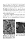
Purchased for the exclusive use of nofirst nolast (unknown)
From: SCC Media Library & Resource Center (library.scconline.org)