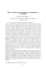
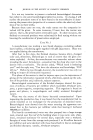
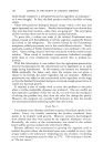
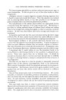
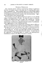
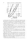
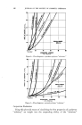
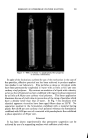
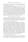
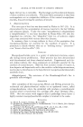
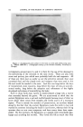
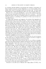
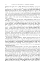
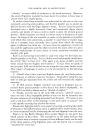
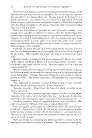
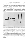
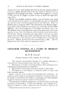
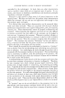
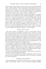
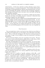
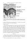
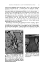
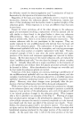
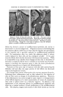
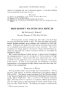
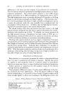
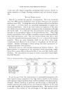
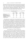
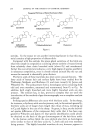
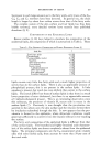
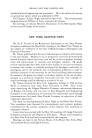
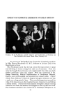
196 JOURNAL OF THE SOCIETY OF COSMETIC CHEMISTS ...... : . •k• • • :•-', ß ,. • . . ,•:'•* .... •...... • •.,.... :.-.•.... " ': . • '-•, •':• .- •* ..• •' -• ..•.• .• . ß • .'•. s• ...•½: :"• .•- . . •., ..... .. -?• •.• :. ........ ,•,, •,,,.-_. ,•. • •½ . .•::: :•: •:. ½•.. •..• .. ...• .. •., . .•: :•½?'•?.. .:..... •:.• .• .. .-...•,: . • . ,.. •..'• ......... :.% •,• •:....._....• •' .'• ..... •T• ' '•" ,.. ,. •.. •-?%. "• :'• • -... •,•, '.., .• ß 4 :..½ Figure 4.--Dermal i,flammatio,: S•alp with prolifera6ve changes after the i,je•tio, of 0.1 •. of 0.1% •roto, oil i, olive oil. (A) (X16'/•). There is marked acanthosis suggesti,• pseudo- epitheliomatous hyperplasia overlyi,g the polymorphous i,•ltr•te a,d •broMastic p•lifera- 6o, i, the dermis. In some ½oci the basal layer is ,ot de•,itely ide,ti•ble where the tory in•ltrate and edeEa is extremely dense and •netrates the overlying epithelium. No mature sebaceous •la.ds •re visible. (•) (X20). This is a d•per sectio, i, the same block of tissue the 1obul•tio,s of the hyperplastic epithelium sug- gests their origi, ,ot o,ly from the follkular ter•al •ot sheath but also from •e sebaceous ½a.d. There is one solitary focus of Eature sebaceous •ells (SC) at the right. ... . .,.• . -,:-:- -. . ':• •' •,•.'.. ..... '-'- Figure $.--Dermal inflammation: Scalp 2 weeks after the intradermal injection of 0.03 cc. of sebum (Xll). Each of the glands (SG) at the top right of the two follicles shown have not only become undifferentiated but have undergone keratinous meta- plasia. Each 1obule is a stratified squamous epithelium producing a comedo-like mass of keratinized squa- dermis often mimicking what the histopathologist is accustomed to calling pseudoepitheliomatous hyperplasia (Fig. 4). The ruptured follicular epithelium forms proliferating sheets and tongues of epithelial cells which grow out in all directions, oftentimes tending to encapsulate the inflam- matory mass. In early specimens some of these proliferating tongues can be seen coming from sebaceous glands. Along with the replacement of the glands and proliferation there are varying degrees of transformation of the

RESPONSE OF SEBACEOUS GLAND TO EXPERIMENTAL STRESS 197 sebaceous glands into stratified squamous epithelium. In other words, the sebaceous gland now instead of forming sebum, forms keratin like any other stratified squamous epithelium and the cavity of the sebaceous gland becomes filled with keratin (Fig. 5). The changes following inflammation are nonspecific and are only dependent upon the degree of inflammation produced. These changes are not dependent on any one specific agent. All of these experiments illustrate the stereotyped response of the se- baceous gland to injury, whether this injury is directed directly at the glands or at the glands as an integrated portion of the pilosebaceous apparatus. The sebaceous cells stop forming their mature product, namely, sebum and form cells which are able to participate in the regenerative response. In instances where these changes have been followed throughout their course, such as after dermabrasion, the changes are reversible and could be easily missed. There is a similarity to the changes seen regularly in hair follicles at the end of each cycle (telogen). In telogen the matrix cells stop forming keratin and form a column of undifferentiated epithelial cells. In respect to the hair the telogen-like pattern occurs regularly after many injuries such as x-ray, injection of colchicine, the administration of thallium, fever, etc. (2). Therefore, the stereotyped defense of both the gland and the hair follicle involves the cessation of synthesis of their highly differentiated products, sebum or hair-keratin, respectively, and replacement by more primitive, undifferentiated epithelial cells. These changes are another example of the pluripotential capabilities of the cells of the epidermis and its appendages (3, 4). In other words, the mature sebaceous glands still maintain the ability under suitable circumstances to produce keratin rather than sebum. Finally, turning to the clinical applications, the contribution of the pilosebaceous apparatus to pseudoepitheliomatous hyperplasia has already been mentioned. More important are the relationship of these changes to the lesions of acne vulgaris. The exact precipitating event in the formation of a comedo is unknown. However, as the comedo forms, a variable degree of undifferentiation develops in the sebaceous glands and the glands undergo atrophy (Fig. 6). At the same time the follicle gradually becomes distended by a mass of keratinous material. As the keratinous material continues to accumulate the sebaceous glands undergo more and more atrophy and in the far advanced comedo no sebaceous glands are present. As after plucking, the atrophy and undifferentiation appear to be a sequelae of follicular hyperkeratosis and the gland is invariably influenced by the marked follicular disturbance with the distention by the horny material. In chloracne, similar changes occur with even greater distention of the follicle by keratin (Fig. 6). A final word of interpretation is necessary. Since a well-formed comedo will have small sebaceous glands, these glands can not be used in studies
Purchased for the exclusive use of nofirst nolast (unknown)
From: SCC Media Library & Resource Center (library.scconline.org)