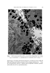
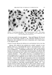
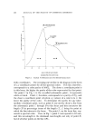
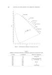
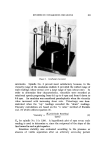
J. Soc. Cosmetic Chemists, 19, 565-580 (Aug. 19, 1968) Melanocytes and Pigmentation* Melanin FUNAN HU, M.D. Presented December 6, 1567, New York 6¾ty Synopsis During their normal development, melanocytes undergo changes in size and shape as well as in enzymatic activitiy. Their developmental phases can be studied in vitro. Young, mature, and old melanocytes are defined according to their morphology, enzyme activity, and ultrastructure. The enzyme tyrosinase that catalyzes the hydroxylation of the melanin precursor tyrosine to dihydroxyphenylalanine and to dopa quinone resides in a minute cytoplasmic structure in melanocytes known as the premelanosome. The biosyn- thesis of melanin is regulated by the availability of free tyrosine as a substrate. the presence of factors that activate tyrosinase, and the presence or absence of inhibitors of tyrosinase. The color of skin depends not so much on the number of mdanocytes in it but on the amount of melanin granules that the melanocytes can synthesize and distribute either in the epidermis or in the dermis. Deviation from normal functioning results in abnormal pigmentation. INTRODUCTION The color of the skin is one of man's major concerns. This area naturally is of great interest to the cosmetic chemists. Skin color varies with the over-all thickness of the integument, the state of vascularity and the amount of the pigment in the skin. Accord- ing to Edwards and Duntley the five primary pigments which contribute to the color of human skin are carotene (yellow), oxyhemoglobin (red), reduced hemoglobin (bluish), melanin, and melanoid (1). Among these, * Publication No. 293 from the Oregon Regional Primate Research Center, supported by Grant FR 00163 of the National Institutes of Health and Grant CA 08499 froin the National Cancer Institute. } Oregon Regional Primary Research Center, 505 N.W. 185th Ave., Beaverton, Ore. 97005.

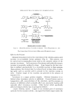
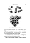
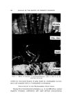
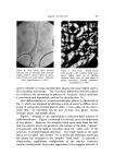
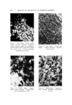
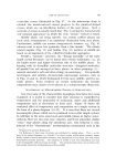
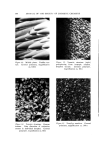
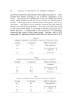
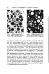
566 JOURNAL OF THE SOCIETY OF COSMETIC CHEMISTS melanin and melanoid are the most important since these are the pig- ments which distinguish the dark-skinned from the light-skinned indi- viduals. It is well established that melanocytes, located in the basal layer of the epidermis, are the only cells capable of melanin production. There- fore, normal or abnormal melanin pigmentation is directly related to anatomical, physiological and biochemical variations of the melanocyte. Hence, when one speaks of melanin pigmentation, one has to speak of the melanocyte. A thorough knowledge of the biological properties of melanocytes is an important prerequisite for the understanding of normal and abnormal pigmentation. Terminology For convenient discussion it is important to define clearly the various terms used in the text. The terminology used here follows that proposed by Fitzpatrick, et al. (2), which is a modification of the one adopted by the Third Con- ference on the Biology of the Normal and Atypical Pigment Cell in 1931. This new version has the approval of the Sixth International Pigment Cell Conference (3). Melanocyte.* A cell which synthesizes a specialized melanin-contain- ing organelle, the melanosome. Melanophore. A type of melanocyte that participates with other chromatophores in the rapid color changes of certain animals by the intracellular displacement (aggregation and dispersion) of melano- somes. g/Ielanoblast. An undifferentiated precursor of the melanoeyte (and melanophore). g/[elanosome. t A discrete melanin-containing organelle in which melanization is complete shown by electron microscopy to be more or less uniformly "electron dense" tyrosinase activity not usually demonstrable. ?remelanosome. All distinctive particulate stages in the maturation of melanosomes, with variable electron density possesses an active tyrosinase system after the onset of melanin synthesis. * Included here are differentiated cells which synthesize nonmelanized or partly melanized premelanosomes as terminal products. It is suggested that in albinism the •nelanoeytes containing nonmelanized premelanosomes be called albino rnelanocytes. • Multiple melanosomes imbedded in supporting matrices (for example, as in the maero- phages and malpighian cells of mammals) may be designated rnelanosorne complexes.
Purchased for the exclusive use of nofirst nolast (unknown)
From: SCC Media Library & Resource Center (library.scconline.org)