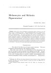
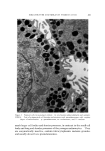
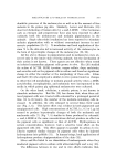
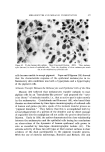
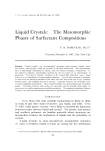
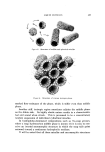
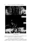
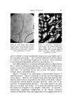
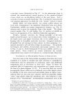
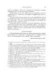
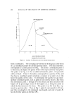
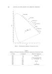
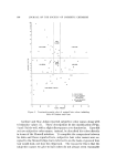
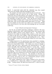
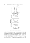
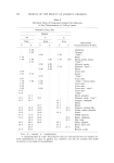
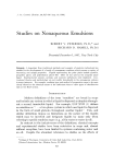
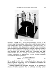
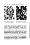
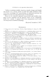
MELANOCYTES AND MELANIN PIGMENTATION 577 i::' 'a o• •' .'?• ,• ' .• : • • ' .::.•,• •---.•.. - •C ' • •. . • .... Figure 10. 21-day human skin culture. cytcs (arrows) iu sheets of epithelial cclls. Note the dcndritcs of these •nclanocytcs reach out to the neighboring epithelial cells cells became unable to accept pigment. Papa and Kligman (38) showed that the characteristic response of the epidermal melanocytes in in- flammatory skin conditions was both a hyperplasia and a hypertrophy of the pigment cells. •,lJelanin Transfer Between the Melanocytes and Epithelial Cells of the Skin Masson (39) believed that melanocytes transfer melanin to mal- pighian cells by an "inoculation-like process" and proposed the "cyto- crine theory" of melanin transfer, i.e., that an active part is taken mostly by the melanocytes. Cruickshank and Harcourt (40), who based their theories on observations by time-lapse cinemicrography of cultured cells of human and guinea pig skin, speak of the melanin transfer process as "pigment donation." They believe that this is accomplished both by actual phagocytosis of a portion of the dendrite and by direct passage of organelles into the malpighian cell not unlike the process described by Masson. Early in 1936, the author demonstrated the close relationship between the melanocytes and the epithelial cells basing her conclusions on observations of the dynamics of human epidermal cells grown in vitro as recorded by time-lapse cinemicrographic technic (41). The extreme activity of these two cell types at their contact surfaces is clear evidence of the dual participation in the pigment transfer process. With the use of electron microscopy, Barnicot and Birbeck (42) and

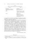
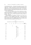
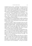
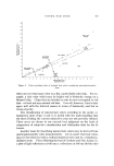
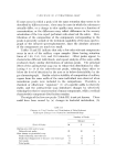
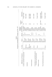
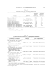
578 JOURNAL OF THE SOCIETY OF COSMETIC CHEMISTS Table II Melanin Transfer and Block in Normal and Abnormal Skins Transfer Block Normal skin Pigmented basal cell epithelioma Pigmented seborrheic keratosis (46) Ephelis Lentigo Vitiligo (30) Precancerous keratosis (45) Melanoacanthoma (46) Inflammatory dermatoses (atopic dermatitis, chronic eczematous dermatitis, Lichen planus) (36, 37) Thorium X (29) Eccrine poroma (47) Epithelium of the intraepider- mal part of sweat duct (47) Outer root sheath epithelium of fetal hair (48) Drochmans (43, 44) showed that segments of melanocytic dendrites containing the pigmented organelles actually penetrate into the mal- pighian cells and are there nipped off. Melanin transfer and block between the melanocytes and malpighian cells occur both in normal and abnormal conditions. Table II lists some examples of these conditions. Hypopigmentation results from the destruction of melanocytes, as in inflammatory conditions of the skin, or from the block of melanin transfer to the epithelial cells in spite of the presence of hypertrophic and hyperplastic melanocytes in the neighboring areas. Conversely, hyperpigmentation results from an in- creased activity of the melanocytes and from increased pigment transfer from the active melanocytes to the epithelial cells. Since in human epidermis the number of the epithelial cells far exceeds that of the melanocytes, and the degree of melanin pigmentation is directly cor- related to the amount of melanin in the epidermis, any change in the amount of melanin in the epithelial cells will influence skin pigmentation more effectively than the change in the melanocytes. (Received December 8, 1967) REFERENCES (1) Edwards, E. A., and Duntley, S. W., Pigment and color of living human hair, Am. J. Anat., 65, 1-33 (1939). (2) Fitzpatrick, T. B., Quevedo, W. C., Levene, A. L., McGovern, V. J., Mishima, ¾., and Oettle, A. G., Terminology of vertebrate melanin-containing calls, Science, 152, 88-89 (1966).
Purchased for the exclusive use of nofirst nolast (unknown)
From: SCC Media Library & Resource Center (library.scconline.org)