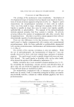
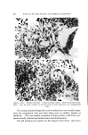
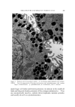
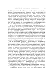
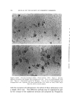
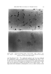
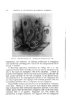
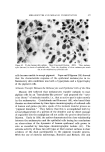
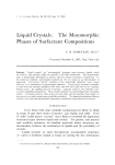
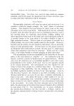
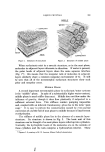
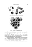
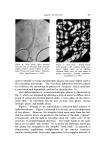
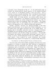
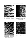
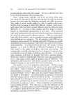
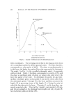
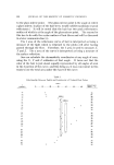
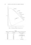
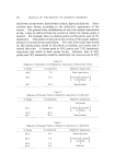
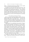
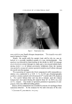
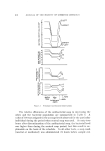
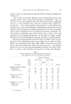
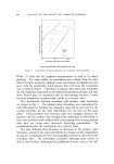
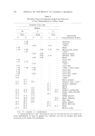
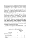
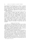
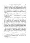
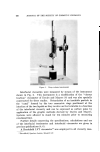
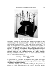
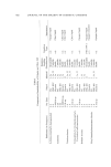
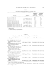
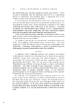
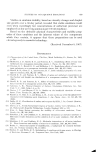
MELANOCYTES AND MELANIN PIGMENTATION 367 CYTOLOGY OF THE MELANOCYTE The cytology of the melanocyte varies remarkably. Description of developmental stages of melanocytes has largely been based on observa- tions of strains of mouse pigment cell grown in vitro (4). Figures 1 and 2 demonstrate the morphological variations of these cells in culture. They vary from small to large although the majority of the cells are bipolar or spindle in shape they may be round, oval, bipolar, spindle, epithelial-like or polydendritic. The amount of the intracytoplasmic melanin pigment granules that they contain is variable. In actively growing cultures the number of nonpigmented cells often exceeds that of pigmented ones. Whether or not they contain pigment these cells are all potentially capable of synthesizing melanin. There are also differences in the fine structure of these cells. The small round or bipolar nonpigmented cells contain only nonmelanized premelanosomes, while the large epithelial or polydendritic pigmented cells contain premelanosomes, melanosomes and melanosome complexes (3, (3) (Fig. 3). The activity of the enzyme, tyrosinase, is also not uniform. With the use of autoradiography, the incorporation of Dopa-2-C TM was not usually observed in those cells that contain large amounts of melanin pigment. The small round, ovoid, or spindle-shaped cells were not regu- larly labeled. The small bipolar or dendritic cells, on the other hand, often showed the uptake of the radioactive substances (7). Similar variations have been recorded in human melanoeytes (8-11). By correlating the morphology, enzyme activity and ultrastructures of these cells, one can divide the pigment cells into young, active, and old. These different forms are included in Table I. The melanoblasts, propigment cells, or immature pigment cells are small, may be round, ovoid, or somewhat triangular in shape, are usually enzymatically inactive, contain no visible melanin pigment, but have premelanosomes. Table I Cytological and Enzymatic Variations in the Melanocytes Propigment Active Old Cell Melanocyte Melanocyte Tyrosinase Premelanosome Melanosome Melanin complex

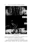
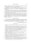
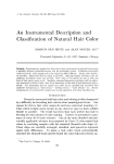
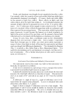
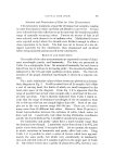
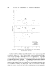
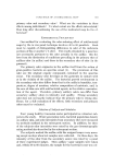
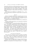
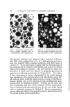
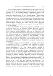
JOURNAL OF THE SOCIETV OF COSMETIC CHEMISTS ...... . . .: •.. :,• . :•- : •. •.• .'••• Figures I and •. Pigment cell strain. 10-day monolayer culture. May-Oruenwald-Oiemsa. 160X. Note the great variation in size and shape of both pigmented and nonpigmented cells The young and physiologically active melanocytes are usually larger than the propigment cells and their shape may be stellate, bipolar or dendritic. They are usually tyrosinase or dopa positive, and their cyto- plasm usually contains premelanosomes and melanosomes. The old melanocytes usually are the largest of the three they have
Purchased for the exclusive use of nofirst nolast (unknown)
From: SCC Media Library & Resource Center (library.scconline.org)