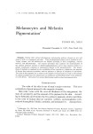
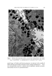
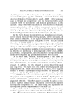
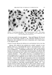
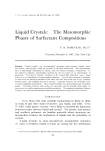
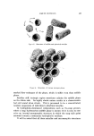
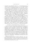
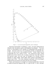
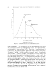
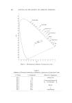
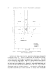
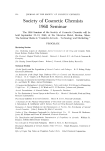
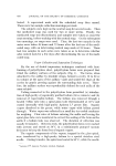
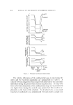
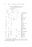
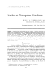
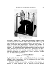
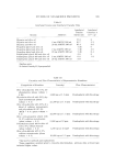
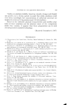
MELANOCYTES AND MELANIN PIGMENTATION 571 /[ TYROSINASE: H l H•, H2 TYROSINE: DOPA Dopoquifiofie (max 280 re.u) HO , z•' HO 0 • COOH HO COOH 5,6 Dlhydraxylfidale DOPACH ROME. Lmacodopachrome (mox 275 298 mr) Red i (max. 305• 475 m•a) Ifidole-5,60ulfiofie ITYRO$1NE:-ME:LANIN I Yellow Browfi { Eumelanlfi) (max 300 450mu) Yellow (Pheomelofilfi) (Generol Absorption) Metabolic pathway of tyrosinc to melanin. Figure {. Mctabolic pathway of tyrosine to melanin. From Fitzpatrick el •l. {49) FACTORS THAT INFLUENCE MELANIN PIGMENTATION 2Ej•ect on the Enzyme Melanin formation involves the conversion of the colorless amino acid, tyrosine, to an insoluble brown polymer (Fig. 4). This process can be carried out in mammalian tissue only by the catalytic action of the enzyme, tyrosinase. In the presence of tyrosinase and molecular oxygen, tyrosine is invariably oxidized to dopa. Dopa formed in the first reac- tion is oxidized enzymatieally by a reversible reaction to dopa-quinone (dopa-quinone thus formed may be reduced back to dopa when a reduc- ing agent such as aseorbie acid is present in the reaction system in vitro). Further stages of the reaction can proceed in the absence of enzymes (16). Tyrosinase is a copper protein complex. Melanin formation is in- hibited when tyrosinase activity is inhibited. General factors such as temperature, pH, concentrations of the substrate, and the presence or absence of inhibitors which normally affect any enzymatic reaction will naturally affect tyrosinase reaction. Other factors which specifically affect tyrosinase activity may be grouped as follows: Copper Binding Agents: Copper is an essential part of tyrosinase molecule for the enzyme activity any agent that binds copper makes the

572 JOURNAL OF THE SOCIETY OF COSMETIC CHEMISTS enzyme inactive. Among these agents, substances possessing a reactive sulfhydryl group will inhibit the enzyme reaction. Early in the 1940's Rothman and co-workers reported the presence of a dialyzable, water- soluble-sulfhydryl-containing component in human epidermis which inhibited the formation of melanin from tyrosine and dopa (17). Hal- prin and Ohkawara (18) have since demonstrated that reduced gluta- thione is the sulfhydryl compound present within epidermal extracts that inhibits melanin formation. They also compared Negro and Cau- casian skins and found that Negro skin contains less reduced glutathione and glutathione reduetase than the Caucasian skin. Adaehi in our Department compared the enzyme activities of the mela•_otic and amelanotie mouse melan.omas and found that the amela- notie tumors have no demonstrable tyrosinase activity but have approx- imately twice the activity of glutathione reduetase as the melanotic tumors (19). Hu, by incorporating agents that have reactive sulfhydryl groups, such as ergothionine and Cleland's reagent, in culture media failed to elicit any inhibiting effects on melanogenesis. At high concentrations these substances inhibit cell growth completely, while lower concentra- tions have 1•o effect whatsoever (7). Reducing Agents: Since the primary reaction involved in the con- version of tyrosine to dopa quinone is an oxidative process, the presence of strong reducing agents will affect this reaction. Ascorbic acid is one example of what is considered to be a depigmenting agent in vitro. When incorporated in the culture medium in which the mouse pigment cells grow, it shows no evidence of inhibiting pigment formation (20). Unknown Tyrosinase Inhibitors: Satoh and Mishima (21) and Chian and Wilgram (22) have demonstrated the presence of tyrosinase inhibitors in Forther's hamster melanoma and $91, B16 and Harding- Passey mouse melanomas. These inhibitors were found in both the pigmented and nonpigmented tumors with higher activity in the latter. Ultraviolet light inaetivates the inhibitor isolated by Chian and Wil- gram. The inhibitor in the Forther's melanoma is not changed by the addition of Cu ++ and therefore does not appear to be a sulfhydryl compound. Effects on the Activity of Melanocytes The effects of hormones, chemicals and radiations have been demon- strated on melanoeytes. Snell (23) reported that a-MSH (melanocyte- stimulating hormone) caused an increase in size and complexity of
Purchased for the exclusive use of nofirst nolast (unknown)
From: SCC Media Library & Resource Center (library.scconline.org)