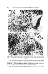
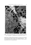
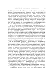
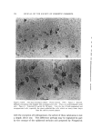
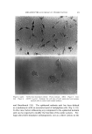
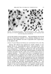
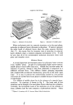
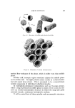
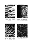
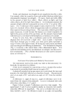
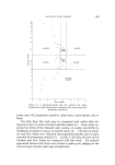
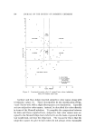
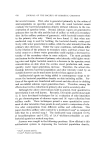
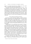
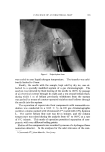
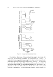
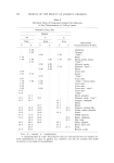
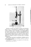
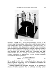
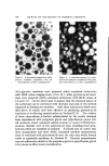
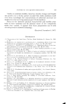
MELANOCYTES AND MELANIN PIGMENTATION 573 dendritic processes of the melanocytes as well as in the amount of free melanin in the guinea pig skin. Similarly, Lerner and McGuire (24) observed darkening of human skin by a and/•-MSH. Other hormones such as estroõen and progesterone have also been reported to affect variously both the melanocytes and melanin pigmentation in the animals. Single ultraviolet irradiation has been reported to stimulate melanin pigmentation with or without concomitant increase in mel- anocyte population (25-7). X-irradiation and local application of tho- rium X to the skin also led to increased activity of the melanocytes in the form of hypertrophic changes of the melanocytes (28, 29). All the above changes in pigmentation and in melanocytes were ob- served only in the intact skin of living animals, but the mechanism of their action is not known. These agents are not effective when tested on isolated mammalian pigment cells grown in vitro. Hu (20) studied the action of ACTH, MSH, tyrosine, copper sulfate, dopa, melatonin, and ascorbic acid on the pigment cells in culture and found no significant change in either the number or the morphology of these cells. Klaus and Snell (30) who employed a similar in vitro system found no changes in either the cell morphology or melanin granule activity when a-MSH, acetylcholine, norepinephrine, and melatonin were incorporated in the media in which guinea pig epidermal melanocytes were cultured. On the other hand, colchicine, a mitotic poison, is not known to stimulate melanocytes. But Hu (20) has shown that, when these cul- tured pigment cells were treated with colcemid for a prolonged period of time (24 to 48 hours), the number of the melanin-containing cells in- creased. In addition, the cells enlarged to several times their usual size (Fig. 5, 6). This latter effect was evident in both pigmented and nonpigmented cells. High concentrations of ACTH, i.e., 0.5 or 5 t•g/ml seemed to produce hypertrophic changes of both the melanotic and amelanotic cells (7) (Fig. 7, 8) similar to those produced by colcemid. a- and/•-MSH at the same concentrations did not produce an effect on the pigment cells as significant as that of ACTH. Hydroquinone in certain concentrations, however, had a selective toxic effect on the pigmented cells (20). This effect appears to parallel its action in vivo. Chavin reported similar changes in pigment cells when he injected hydroquinone into goldfish (31). In human beings, local applications of hydroquinone produce depigmentation of the skin (32). Silver and Hu failed to see stimulation of melanogenesis when they irradiated pigment cells in culture with ultraviolet light and x-ray (33). The difference between in vivo and in vitro effects indicates that,

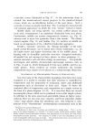
574 JOURNAL OF THE SOCIETY OF COSMETIC CHEMISTS Figures 5 and 6. Nine-day monolaycr culture. Phase contrast. 140X. Figure 5. Control. Melanin-coutaining cells mingled with nonpigmented cells. These are predominantly small cells. Figure 6. Colcemid 0.3 vg/ml for 48 hours. Note the size of both pigmented and nonpigmented cells, especially the giant epithelial-like cells, which are many times larger than the cells in control cultures with the exception of hydroquinone, the action of these substances is not a simple, direct one. This difference perhaps may be explained in part by the concept of the epidermal melanin unit proposed by Fitzpatrick
Purchased for the exclusive use of nofirst nolast (unknown)
From: SCC Media Library & Resource Center (library.scconline.org)