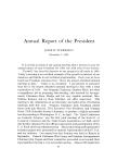
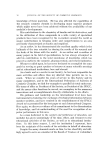
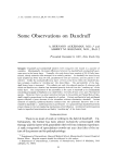
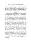
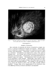
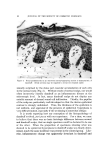
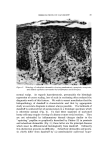
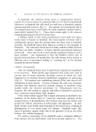
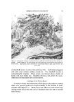
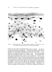
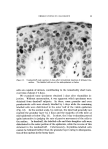
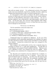
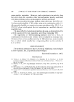
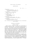
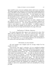
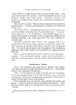
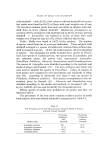
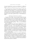
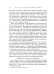
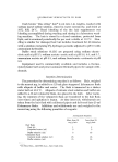
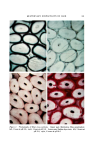
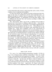
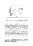
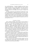
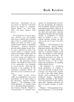
86 JOURNAL OF THE SOCIETY OF COSMETIC CHEMISTS Figure 4. Focal parakeratosis in an otherwise normal-appearing section is characteristic of dandruff. Serial sections may be required to reveal the retained nuclei sionally surprised by the dense peri-vascular accumulation of such cells in the normal scalp (Fig. 3). Without study of normal scalps, one would often incorrectly classify dandruff as an inflammatory disease at the microscopic level. In fact, many dandruff scalps do not display any notable amount of dermal round cell infiltration. Thirdly, the papillae of the scalp are particularly well-developed so that the dermo-epidermal contour is strongly undulated. Thus, the thickness of the epidermis is not uniform, and appraisal of the presence of epidermal hyperplasia is very difficult indeed, especially if the sectioning is somewhat oblique. It .is worth reporting how our conception of the histopathology of dandruff evolved, pari passu with our experience. For a time, we came to believe that there was no basic histologic difference between normal and dandruff scalps, that no single specimen could be declared to be one or the other. When the proportion of specimens showing what was deemed to be epidermal hyperplasia in dandruff scalps was finally deter- mined, much the same incidence was present in the control group. Like- wise, inflammatory change was apparently invariant in dandruff and

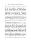
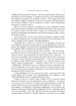
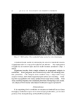
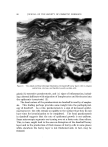
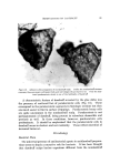
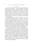
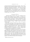
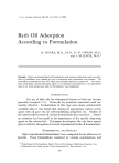
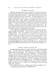
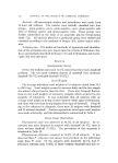
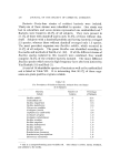
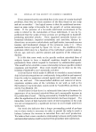
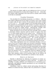
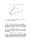
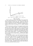
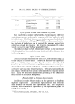
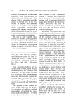
OBSERVATIONS ON DANDRUFF 87 Figure 5. Histology of seborrheic dermatitis showing parakeratosis, spongiosis, exocytosis, and dilated capillaries surrounded by lymphocytes and histiocytes normal scalps. As regards hyperkeratosis, presumably the histologic expression of excess scaling, loss of scale in sectioning often reduced the diagnostic worth of this feature. We wish to assert nonetheless that the histopathology of dandruff is characteristic and that by appropriate study an accurate diagnosis is almost always possible. The hallmark of dandruff is scattered loci of parakeratosis in a histologic specimen which is otherwise normal (Fig. 4). To find these segments of nucleated horny cells may require viewing of a dozen or more serial sections. They are not subtended by inflammatory dermal changes similar to the "squirting" papillae so graphically described by Pinkus (2) in psoriasis and seborrheic dermatitis (Fig. 5) these latter are the principal diseases which must be differentiated histologically from dandruff. Ordinarily this distinction presents no difficulty. Seborrheic dermatitis and psoria- sis chiefly differ from dandruff by (a) unmistakable epidermal hyper-
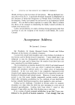
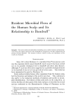
Purchased for the exclusive use of nofirst nolast (unknown)
From: SCC Media Library & Resource Center (library.scconline.org)