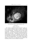
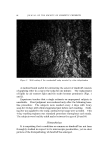
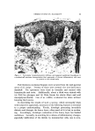
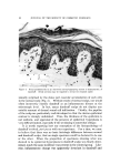
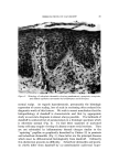
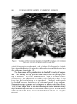
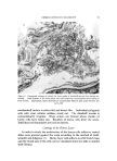
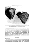
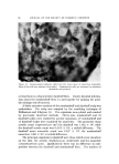
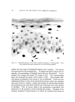
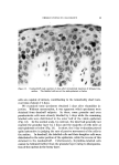
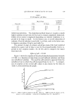
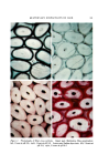
lOO JOURNAL OF THE SOCIETY OF COSMETIC CHEMISTS DISCUSSION From the preceding studies a tentative conception of the nature of dandruff has been formulated. It is proposed that profuse scaling of the scalp, the hallmark of dandruff, represents a fundamental alteration in the rate regulating mechanism of horny cell production, i.e., epidermal turnover is increased. The rapid transit of cells to the surface does not allow time for complete keratinization. This is betrayed by the presence of parakeratotic cells. Horny layer coherence is affected and cracks occur deep in the horny layer, producing large flakes. The intensified scaling reflects both a true increase in the production of horny cells and their being rendered more conspicuous by "cracking up" and desquamat- ing as large aggregates. In dandruff, unlike other conditions in which there is an increased turnover rate of the epidermis, there is no histologic evidence of an antecedent inflammatory stimulus to power the process. Nor is there any other characteristic histologic change in the living part of the skin. The principal pathology is located in the horny layer. The dead horny layer cells must reflect changes that occurred when they were living and made their upward migration. These aberrations in the living epidermis and perhaps the dermis are recondite and will require the more powerful tools of electronmicroscopy and biochemistry to define them. McOsker and Hannon (5), in their electron microscopic studies, did show striking alterations in the stratum corneum, but not in the viable epidermis. It is therefore postulated that changes in the dandruff horny layer, the chaotic pattern, "cracking up" into large cellular aggregates, crev- ices, and sporadic parakeratosis, are secondary to more rapid epider- mopoiesis and perhaps some subtle change in epidermal physiology. Whenever the rate of keratinization is accelerated, there is faulty cohe- sion and evidence of incomplete keratinization. In terms of epidermal kinetics, dandruff is perhaps intermediate between normal skin and frankly pathologic conditions such as seborrheic dermatitis and psoriasis. If the structural and chemical changes characteristic of dandruff are subsequent to increased epidermopoiesis, one might anticipate that the biochemical changes in the dandruff horny layer would correspond to those in psoriasis but to a far lesser extent. Preliminary data suggest that these changes do occur in a diminutive way (15, 16). Dandruff scales contain a high sulfhydryl and pentose content and lower than nor- mal amounts of free amino nitrogen.

OBSERVATIONS ON DANDRUFF lol ACKNOWLEDGMENTS The technical assistance of Miss Rosalin Bloomberg is gratefully acknowledged. Analysis of organisms on dandruff and nondandruff scalps was carried out by Mr. Peter Williamson and Dr. Richard Mar- ples. Inmates of Holmesburg Prison, Philadelphia, served as volunteers for this study, and Edward Hendrick, Superintendent, gave permission for use of the prison facilities. Mr. Edward Gliffort kindly did the photography. (Received April 16, 1968) REFERENCES (1) Goldschmidt, H., and Kligman, A. M., Quantitative estimation of keratin production by the epidermis, Arch. Dermatol., 88, 709-12 (Dec. 1963). (2) Pinkus, H., and Mehregan, A. H., The primary histologic lesion of seborrheic dermatitis and psoriasis, J. Invest. Dermatol., 46, 109-16 (Jan. 1966). (3) Kligman, A.M., and Christophers, E., Preparation of isolated sheets of human stratum corneum, Arch. Dermatol., 88, 702-5 (Dec. 1963). (4) Christophers, E., and Kligman, A.M., Visualization of the cell layers of the stratum comeurn, J. Invest. ])ermatol., 42, 407-10 (1964). (5) McOsker, D. E., and Harmon, D. P., Ultrastructural studies of dandruff-involved scalp tissue, Proc. Sci. Sect. Toilet Goods Assoc. No. 4, 5-7 (May 1967). (6) Facq, J. D., Kirk, L., and Rebell, G., A simple replica technique for observation of human skin, J. Soc. Cosmetic Chemists 15, 87 (1964). (7) Goldschmidt, H., and Kligman, A.M., Exfoliative cytology of human horny layer, Arch. Dermatol., 96, 572-6 (Nov. 1967). (8) Williamson, P., in Maibach, H. I., and Hildick-Smith, G., eds., Skin Bacteria and Their Role in infection, McGraw-Hill, New York, N. Y., 1965. (9) VanderWyk, R. W., and Roia, F. C., The relationship between dandruff and the micro- bial flora of the human scalp, J. Soc. Cosmetic Chemists, 15, 761-8 (1964). (10) Sabouraud, R., De la seborrhea iVouvelle Pratique Dermatologique, Vol. 7, Masson et Cie, Paris, 1936, pp. 1-60. (11) Marplcs, M. J., The Ecology of the Hutnan Skin, Charles C Thomas, Springfield, II1., 1965. (12) Marpies, R. R., and Williamson, P., Milliore filter method for quantitating Pityro- spoturn, unpublished data. (13) Weinstein, G. D., and Van Scott, E. J., Autoradiographic analysis of turnover times of normal and psoriatic epidermis, J. Invest. Dermatol., 45, 257-62 (October 1965). (14) Epstein, W. L., and Maibach, H. I., Cell renewal in human epidermis, Arch. Dermatol., 92, 462-8 (October 1965). (15) Wheatley, V. R., Flesch, P., Esoda, E. C., Coon, W. M., and Mandol, L., Studies of the chemical composition of the horny layer lipids, J. invest. Dermatol., 43, 395-405 ( Nov. 1964). (16) Laden, K., A comparative chemical study of dandruff flakes, skin scrapings, and callus, J. Soc. Cosmetic Chemists, 16, 491-7 (1965).
Purchased for the exclusive use of nofirst nolast (unknown)
From: SCC Media Library & Resource Center (library.scconline.org)