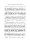
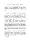
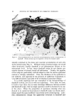
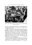
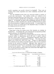
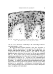
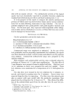
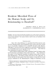
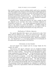
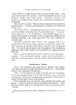
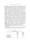
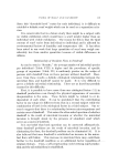
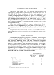
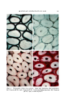
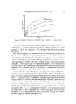
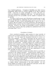
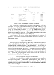
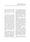
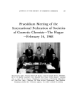
98 JOURNAL OF THE SOCIETY OF COSMETIC CHEMISTS Figure 12. Dandruff specimen 5 days after intradermal injection of •H-thymidine. Most of the labelled cells have reached the top of the living epidermis midinc into the scalps of 6 dandruff subjects and 5 normals. The tissues were processed in the standard way. In biopsy specimens taken after 45 minutes, the percentage of labelled basal cells was determined. In the normal, the average was about 7% (range ,5-9%). The corresponding figure for dandruff was about 13% (range 8-19%). These figures indi- cate almost a doubling of the turnover rate in dandruff. There was one additional finding in some of the dandruff specimens which also signified increased turnover, namely, a higher percentage of suprabasalar labelled cells indicating that cells which had left the basal layer were still capable of dividing. This, of course, adds to the germinative population and ultimately to the quantity of cells reaching the surface. Psoriasis is the ultimate example of rapid turnover in which the bottom three layers of

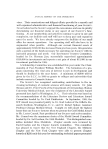
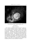
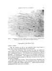
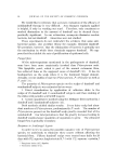
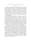
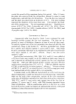
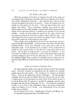
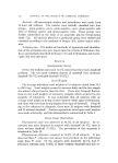
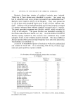
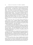
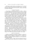
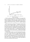
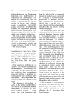
OBSERVATION:S ON DANDRUFF Figure 13. Nondandruff scalp specimen 5 days after intradermal injection of tritiated thy- midine. The labelled cells are at the mid-epidermis or below cells are capable of mitosis, contributing to the remarkably short turn- over time of about 3-4 days. We examined some specimens obtained $ days after thymidine in- jection. Without mensuration, it was apparent which specimens were obtained from dandruff subjects. In these, some granular and even parakeratotic cells were already labelled by $ days while the remaining labelled cells were distributed in the outer half of the viable epidermis (Fig. 12). In the normal scalp, by contrast, the label had generally not reached the granular layer by 5 days and the majority of cells were at mid-epidermis or below (Fig. 13). In short, the 5-day evaluation proved quite instructive in judging the rate of passive movement of the cells to the surface. In dandruff, the labelled cells and their daughter cells were distributed in the outer portion of the epidermis, while the reverse of this obtained in the nondandruff. Unfortunately, thymidine-labelled cells cannot be followed further than the granular layer owing to disorganiza- tion of the nucleus in the horny layer.
Purchased for the exclusive use of nofirst nolast (unknown)
From: SCC Media Library & Resource Center (library.scconline.org)