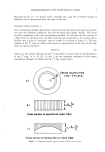
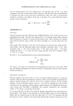
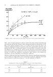
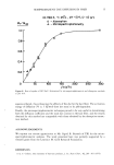
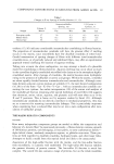
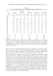
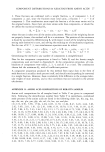
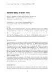
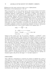
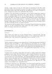
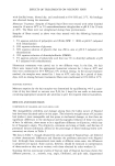
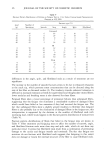
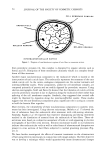
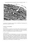
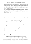
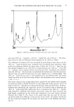
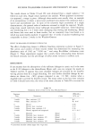
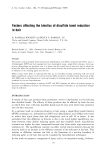
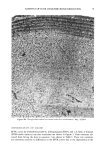
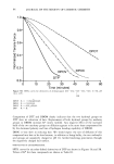
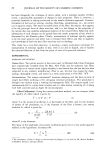
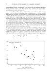
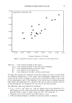
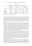
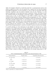
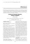
j. Soc. Cosmet. Chem., 36, 53-59 (January/February 1985) Electron microscopy-image analysis: Quantification of ultrastructural changes in hair fiber cross sections as a result of cosmetic treatment JACALYN G. GOULD and RAYMOND L. SNEATH, The Gillette Company, Personal Care Division, Gillette Park, Boston, Massachusetts 02106. Received October 31, 1984. Presented at the Society of Cosmetic Chemists Annual Meeting, New York, December 6-7, 1984. Synopsis In a recent publication, Kaplin et al. described the dissolution of ultrastructural components in hair fibers as a direct result of various cosmetic treatments. These observations were based on a subjective assessment of holes or voids observed in electron micrographs of hair fiber cross sections. We have further investigated these effects using electron microscopy in conjunction with image analysis. Cross sections of proximal and distal ends of intact hair fibers were examined before and after repeated shampooings. The total number of holes, total projected areas, mean areas, and size distributions were determined in the cuticle and cortex regions of hair fibers using a Quantimet 900 Image Analyzer. All measured parameters indicated that ultrastructural disruption increased from the proximal to the distal end of hair fibers. The-impact of shampooing was limited to the cuticle region of weathered hair fibers. INTRODUCTION The ultrastructural organziation of the hair fiber has been extensively studied over the past decade. The keratin components of the hair, which constitute about 85%'of the mass of the fiber, are considered to make the major contribution to the chemical and physical properties of the hair. The remainder of the fiber consists of non-keratinous materials in the form of intercellular membranes and the remnants of cellular constit- uents. These non-keratinous materials have recently been the subject of investigations concerning their contribution to the integrity of the fully keratinized hair fiber. Figure 1 provides a schematic diagram of the non-keratinous regions of the hair fiber in transverse section. The detailed structure and chemical composition of these regions have been revealed by electron microscopy used in combination with various chemical studies. The intercellular membranes form a network structure between both cuticle and cortical cells. Electron micrographs show that these membranes consist of two lipid layers, the [3-1ayers, and a central non-keratinous protein layer, the 8-layer, often referred to as the intercellular cement (1-3). The cell membranes and the intercellular cement make up the so-called cell membrane complex of keratin fibers. Although resistant to attack 53

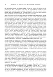
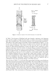
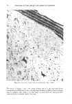
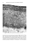
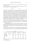
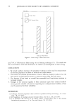
54 JOURNAL OF THE SOCIETY OF COSMETIC CHEMISTS CELL MEMBRANE COMPLEX ENDOCUTICLE • ß NUCLEAR REMNANT ß • ß MELANIN GRANULE INTERMACROFIBRILLAR MATRIX Figure 1. Diagram of non-keratinous regions of hair fiber in transverse section. from proteolytic enzymes (4), this complex is disrupted by organic solvents such as formic acid (5). Disruption of these membranes ultimately results in a complete break- down of fiber structure. Another major non-keratinous component is the endocuticle which is located at the inner portion of each cuticle layer. The endocuticle represents the remnants of the once viable cuticle cell. In the cortex, analogous components are the nuclear remnants and intermacrofibrillar matrix. These components, unlike the cell membrane complex, are composed primarily of protein and are readily digested by proteolytic enzymes. Using electron micrographs, Swift and Bews (6) showed that the liberation of cuticle cell-like units by proteolytic enzymes is due to digestion along the endocuticle sheet rather than splitting of the cell membrane complex. Similarly, they suggested that cortical cells might be liberated by digestion along the intermacrofibrillar matrix. These studies suggest that the non-keratinous components play a significant role in acting as a cement to hold the keratin fiber together. More recently, the vulnerability of these non-keratinous components to cosmetic treat- ments has been investigated. Using electron microscopy, Mahrle eta/. (7) showed the loss of substances from the endocuticle after treatment with cold-waving solutions. Similarly, Kaplin eta/. (8) reported that repetitive shampooing and drying treatments resulted in the dissolution of material from the endocuticle of hair fibers. These ob- servations were based on a subjective assessment of holes or voids appearing in electron micrographs of hair fiber cross sections. Electron microscope studies in our laboratory indicate that endocuticular voids as well as other forms of ultrastructural disruption are present in cross sections of hair fibers subjected to normal grooming processes (Fig- ure 2). We have further investigated the effects of cosmetic treatments on the ultrastructure of hair using electron microscopy in conjunction with image analysis. Our first objective was to develop a technique which would quantitatively assess ultrastructural changes
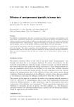
Purchased for the exclusive use of nofirst nolast (unknown)
From: SCC Media Library & Resource Center (library.scconline.org)