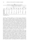
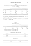
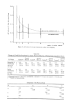
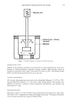
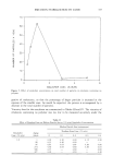
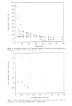
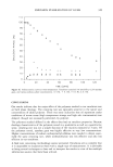
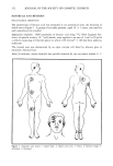
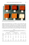
CLEANSING BAR EVALUATION 311 (10), fluorescence enhancement by humidity (11), and phosphorescence quenching (of tryptophan) (11) by humidity. b) The range of fluorescence of NADH (12). The concentration of NADH is decreased by inflammatory processes. The cleansing efficacy of products may be evaluated by fluorometric analysis of the residues of (extrinsic) skin-contaminating fluorophors after cleansing (8). The skin-care potential of products may be estimated by taking into account several parameters characterizing the structure of the horny layer: a) Skin profile measurements demonstrate objectively the evenness of the horny layer (13). The visual inspection of skin sites after patch testing includes the evaluation of fissures (14). b) Trans-epidermal water loss reflects the structural integrity of the horny layer (15). EXPERIMENTAL MATERIALS The following commercially available soaps were used as 2% and 8% solutions in dis- tilled water: A (surfactant-type soap, containing sodium-cocyl-isethionate-tallowate and isethianote etc.) B (classical soap) C (classical soap, containing amines as neutralizing agents) D (classical soap, sodium salts of fatty acids + additional lipids) EPICUTANEOUS TEST Visualscoring andskin surfacepH values. Twenty volunteers (10 m, 10 f average age • = 33.2 years s = 12.3) took part in the study performed December 3-11, 1984. The volunteers had healthy normal skin. Informed consent was obtained. 100 ptl of the two solutions (40øC) of each product were transferred to filter paper discs (diameter = 11 ram) fitted to Finn chambers (diameter = 11 ram manufacturer, Epitest Ltd. Oy, Helsinki, Finland). The chambers were fixed by Scanpor (Norge- plaster, Oslo, Norway) to the volunteers' backs 4 cm to the left and right of the spine. The products were applied in a randomized manner. The different dilutions were ap- plied at contralateral sites. An empty (control) chamber was also fixed within each column. Twenty-four hours after application the chambers were removed. Twenty-four hours after chamber removal the irritation was evaluated using the following scale: 0 = no reaction. 0.5 = weak erythema. 1 -- strong erythema. 1.5 = strong erythema, blisters, fissures. Immediately after removal of the chambers, the pH values of the residual solutions at the skin surface were measured by means of a conventional pH electrode.

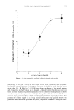
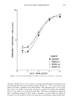
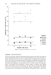
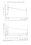
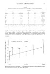
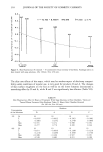
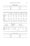
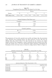
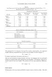
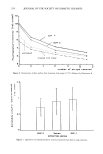
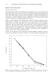
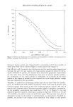
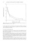
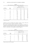
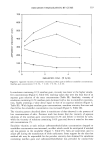
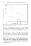
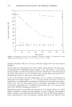
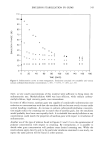
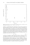
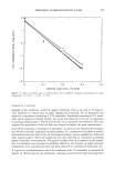
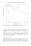
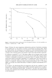
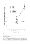
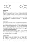
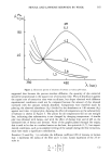
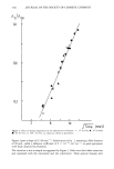
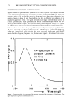
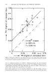
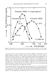
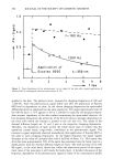
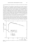
312 JOURNAL OF THE SOCIETY OF COSMETIC CHEMISTS Measurement of skin surface fluorescence. Twenty-four hours after removal of the Finn chambers the skin surface fluorescence of the spots which had been occluded for 24 hours and of neighboring untreated areas was determined. A bifurcated quartz fiber optic (diameter = 4 mm) (Schott, Mainz, FRG) was used. This Y-shaped device was connected to the excitation and emission window of a fluorometer (LS 5, Perkin Elmer). The end, where the single fibers are intermingled, was placed perpendicular to the skin at a distance of 2 mm. The working conditions were a) Excitation 290 nm, emission 320-500 nm. b) Excitation 360 nm, emission 400-500 nm. The spectra were recorded, stored, and finally converted to an average spectrum by computer (type 3600, Perkin Elmer). Evaluation of cleansing efficacy. Skin sites (diameter = 1.5 cm) on the inner side of the lower forearm were contaminated with 1 mg mineral oil/cm 2 containing 0.1% anthra- cene which can be determined by fluorimetry (excitation, 353 nm emission, 400 nm) the instrumentation was the same as described above. Informed consent was obtained. The fluorescence of the skin surface was determined before contamination (= F•) and three minutes after contamination (= F2). The skin sites were cleaned with soap and warm water (32 ø C, 30 sec., regular use). Then the sites were dried by slight touching with a towel. One minute later the residual fluorescence (= F3) was determined. The relative contamination-index was defined as: Rr __ F 3 - F• F 2 -- F• After finishing the measurement, any residue of the fluorophor was removed by .tape stripping. Relative density ofanionic skin surface charge. Ten male volunteers (average age • = 34.5 s = 11.3) took part. Informed consent was obtained. The volunteers washed their lower forearms for one minute on two consecutive days with two of the soaps, using a different soap on each forearm. The soaps were randomly distributed. Two minutes, twenty minutes and three hours after product application, spots (diameter = 3.3 cm) of the treated skin sites were colored with solutions of the cationic dye Rhodamine B (20 ppm in glycerol/H20 = 1'1) for 1 minute. The excess Rhodamine B solution was removed by distilled water washing and the sites dried. By means of quartz fiber optics the fluorescence (Ex 552 nm, Em 590 nm) of Rhodamine B was determined five times at every colored spot. The average values were used for calculation. After finishing the measurement, the color was removed by washing and tape stripping. Skin penetration of soaps D and A. Twenty volunteers (average age • = 31.6 s = 11.1) took part. Funnels containing 0.75% solutions of D and A, respectively, were pressed for two minutes on the lower forearms. Residues of the solutions on the skin surface were wiped off with cotton. An analogous treatment with pure water was performed at one of the upper forearms. The contralateral site remained untreated. Thirty minutes after application, Rhodamine B solution was applied and determined as described above, before and after removal of two, six and eleven tape strips.
Purchased for the exclusive use of nofirst nolast (unknown)
From: SCC Media Library & Resource Center (library.scconline.org)