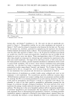
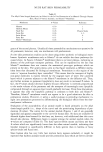
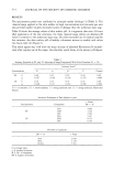
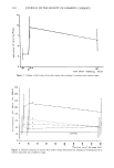
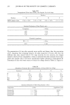
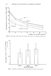
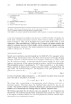
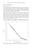
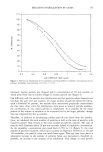
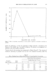
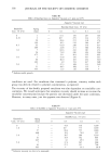
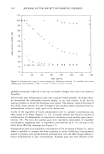
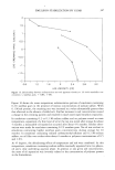
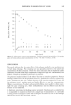
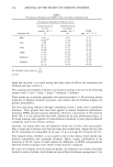
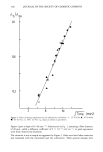
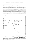
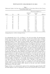
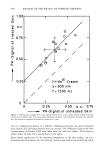
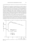
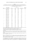
392 JOURNAL OF THE SOCIETY OF COSMETIC CHEMISTS For this reason, we tried to test the direct influence of UV light as a well-standardized irritation source on the possible alteration in acid phosphatase activity. However, it is well documented in the literature that UV light is related to human skin cancer devel- opment (16). Therefore, we switched to an in vitro system consisting of UV light from a germicidal lamp and human skin fibroblasts in culture. In Table III acid phosphatase activities of these cells are shown 0-9 days after irradiation with fluences of 0-45 J/m -2 at 254-nm light. A slight fluence-independent increase in this enzyme activity could be found at day 7. This enhancement is probably due to overall higher protein content. No fluence-response for acid phosphatase activity was detected even nine days after irradiation in our in vitro experiments. It is conceivable that several UV treatments are necessary to stimulate the activity of acid phosphatase. Nevertheless, our results support the notion that UV light is not directly involved in the long-term increase of acid phosphatase activity. Hence, the long-term effects detected in vivo are probably due to indirect mechanisms. In this respect, Prottey et al. (8) found higher acid phosphatase activity as a function of depth of stripping. Therefore, a stimulation of the stratum corneum turnover rate following irritation of human skin could be the cause of the long-term enzyme increase. It is also conceivable that preformed acid phosphatases were released by surfactants or UV light from latent sources as suggested by Rutherford and Table III Acid Phosphatase Activity in Normal Human Fibroblasts Following UV Irradiation (mU/Petri Dish a) Days Following Irradiation a 0 1 2 3 4 7 8 9 Content of protein per Petri dish (tag) b -- 435 455 477 422 662 -- --- 210 ñ 806 ñ 137 ñ 60 + 215 Fluence (J/m2) c 0.0 2.11 1.82 1.44 1.98 2.09 3.44 2.64 --- 0.10 __+ 0.52 ñ 0.03 __ 0.08 1.5 2.48 1.91 2.18 2.04 3.02 3.32 2.12 ___ 0.05 ñ 0.33 3.0 2.43 1.85 1.76 2.11 3.64 3.42 2.30 ñ 0.37 ñ 0.08 6.0 2.15 1.83 1.66 2.23 2.51 4.24 2.19 ñ 0.44 ñ 0.25 ñ 0.7 ñ 0.16 12.0 2.43 1.91 1.69 1.83 2.17 4.05 2.13 ñ 0.20 ñ 0.31 ñ 0.72 ñ 0.06 23.0 -- 1.98 -- -- 1.78 -- -- ñ 0.01 ñ 0.12 45.0 -- 1.81 -- -- 2.02 -- ñ 0.02 ñ 0.22 2.91 ñ 0.26 2.69 3.01 ñ 0.07 3.11 ñ 0.06 3.09 ñ 0.08 a Period following UV irradiation after which acid phosphatase activity was determined. b Content of protein in Petri dish determined after the method of Lowry et al. (11). c Irradiation with 254-nm UV light was carried out as described in Materials and Methods. a Mean values of duplicates are given. In experiments following one and four days after UV irradiation, duplicates of two independent experiments are listed.

SKIN ACID PHOSPHATASE AS IRRITATION MARKER 393 Pawlowski (7). In the present investigation, however, this problem could not be re- solved definitely and requires further studies. ACKNOWLEDGEMENTS We would like to thank Martine Pfefferli, Chantal Michaud, and Eveline Buntschu for skillful technical assistance. Especially, we are indebted to Dr. Thomas C. Brown for critical reading of the manuscript. REFERENCES (1) A. B. G. Lansdown, Appraisal of methods for detecting primary skin irritants, J. Soc. Cosmet. Chem., 23, 739-772 (1972). (2) C. Protrey, "The Molecular Basis of Skin Irritation," in Cosmetic Science, M. M. Breuer, Ed. (Aca- demic Press, London, 1979), Vol. 1, pp 275-349. (3) P. J. Frosch and A.M. Kligman, The soap chamber test,J. Am. Acad. Dermatol., 1, 35-41 (1979). (4) P. J. Frosch, "Irritancy of Soaps and Detergent Bars," in The Principles of Cosmetics for the Dermatologist, Ph. Frost and St. Horwitz, Eds. (Mosby, St. Louis, 1982), pp 5-12. (5) J. E. Wahlberg, Assessment of skin irritancy: Measurement of skin fold thickness, Contact Dermatitis, 9, 21-26 (1983). (6) W. V. R. Shellow and M.J. Rapaport, Comparison testing of soap irritancy using aluminium chamber and standard patch methods, Contact Dermatitis, 7, 77-79 (1981). (7) T. Rutherford and A. Pawlowski, Acid phosphatase staining of the stratum corneum as a marker of damage by low irritancy compounds, Br. J. Dermatol., 91, 503-506 (1974). (8) C. Protrey, D. Oliver, and A. C. Coxon, Prediction and measurement of surfactant action upon human skin under realistic conditions, Int. J. Cosmet. Science, 6, 263-273 (1984). (9) K. Walter and C. Schiitt, "Saure und alkalische Phosphatase im Serum," in Methoden der enzymatischen Analyse, H. J. Bergmeyer, Ed. (Verlag Chemie, Weinheim, 1970), Vol. 1, pp 818-822. (10) H. J. Niggli and P. A. Cerutti, Cyclobutane-type pyrimidine photodimer formation and excision in human skin fibroblasts after irradiation with 313-nm ultraviolet light, Biochem., 22, 1390-1395 (1983). (11) O. H. Lowry, N.J. Rosebrough, A. L. Farr, and R. J. Randall, Protein measurement with the folin phenol reagent, J. Biol. Chem., 193, 265-275 (1951). (12) W. T. Gibson and M. R. Teall, Interactions of C•2 surfactants with the skin: Studies on enzyme release and percutaneous absorption in vitro, Food Chem. Toxicol., 21, 581-586 (1983). (13) G. Hiibscher and G. R. West, Specific assays of some phosphatases in subcellular fractions of small intestinal mucosa, Nature (London) 205, 799 (1965). (14) P. G. M. Van der Valk, J.P. Nater, and E. Bleumink, Skin irritancy of surfactants as assessed by water vapor loss measurements,J. Invest. Dermatol.. 82, 291-293 (1984). (15) F. Stenback, Health hazards from ultraviolet irradiation, Public Health Rev., 10, 229-237 (1982). (16) F. Urbach, J. H. Epstein, and P. D. Forbes, "Ultraviolet Carcinogenesis: Experimental, Global and Genetic Aspects," in Sunlight andMan, T. B. Fitzpatrick, M. A. Pathak, L. C. Harber, M. Seiji, and A. Kukita, Eds. (University of Tokyo Press, 1974), pp 259-283.
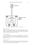
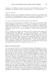
Purchased for the exclusive use of nofirst nolast (unknown)
From: SCC Media Library & Resource Center (library.scconline.org)