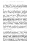
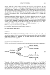
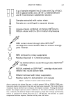
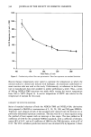
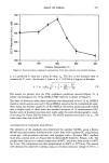
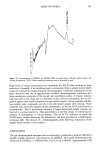
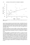
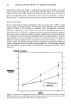
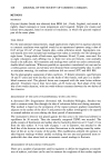
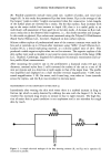
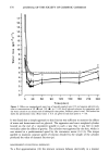
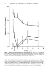
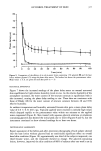
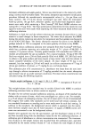
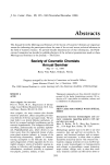
372 JOURNAL OF THE SOCIETY OF COSMETIC CHEMISTS 30- :I, 20- E o 'o10_ 0 Ill 0 2 3 4 5 6 Time ( h ) Figure 4. Reduced skin surface roughness after topical treatment with water (O) (p 0.05 at ten minutes, Student's paired t-test), and 10% aqueous glycerol solution (i,), (p 0.02 at 30 minutes, one hour, and two hours, Student's paired t-test). The broken line describes the change in the roughness of untreated skin sites. Mean values _ S.E. are given for each data point (n = 6). meter which measured a combination of capacitance and resistance was used. The in- strument was developed in conjunction with the Torry Research Station (16). A 2 KHz signal was applied to the skin via a four-terminal device. The delay (in microseconds, }xs) between the applied voltage cycle and the cycle of current flow was measured. The increase in phase delay readings after treatment gave a measure of skin hydration.

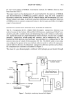
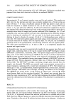
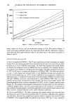
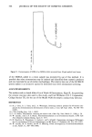
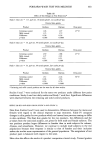
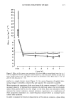
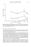
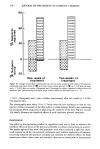
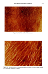
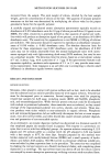
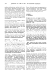
GLYCEROL TREATMENT OF SKIN 373 30- :t. 20- E .c_. .o_ -o 10- 0 - I I I I I I •.•4" -"' I 0 I 2 2__ ____ 4... ____..-•--"'- 6 7 8 24 '"-" '"' Time ( h ) Figure 5. Reduction in skin surface roughness up to 24 hours after treatment with a 15% glycerol oil-in- water lotion (I) and its base lotion without glycerol, (O) (p 0.02 at two hours and five hours, $tudent's paired t-test). The broken line describes changes in the roughness of untreated skin sites. Mean values -+ S.E. are given for each data point (n = 8). PHOTOGRAPHIC ASSESSMENT OF SKIN CONDITION Visual assessment of skin condition is one of the most important parameters by which the consumer assesses the efficacy of a skin care preparation. Studies using experienced observers have been frequently reported (17). In this work macrophotographs (magnification 1:1) of the back of the hand were taken from 15 subjects before and after each treatment period, using Kodak Ektachrome 160 colour reversal film. An Olympus OM1 camera was fitted with a 50-mm macrolens and a 36-mm extension tube and mounted vertically on a copying stand. Lighting was standardised using two opposed 100 W reflector lamps set at an angle of approximately 30 ø to the surface of the skin. The hand was placed in a plaster mould which was clamped to the base of the stand so that the illumination and camera position remained constant relative to the area of skin being photographed. Simultaneous examination of the photographs taken before and after treatment for each subject was made by a team of experienced observers. The difference in overall skin condition was assessed using a better/worse/no difference scoring system. Parameters considered included the depth and frequency of the lines, degree of dryness or flakiness,
Purchased for the exclusive use of nofirst nolast (unknown)
From: SCC Media Library & Resource Center (library.scconline.org)