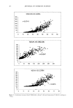
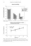
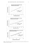
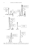
PHOTOSTABILITY OF UV FILTERS 3 Ba/a,ce. All the weighings were done on a Mettler balance (Max 205 g/62 g, d = 0.1 mg/0.01 mg). Syringe. The sunscreen was applied with a 50-pl micro-syringe (Hamilton Bonaduz Schweiz, #705) and spread with a finger glove (Romed, latex finger cots prepowdered, large 4). Adhesive. Tape strippings were done using adhesive tape (Tesa film no. 5529). They were conserved after withdrawal in 10-ml amber flasks (Merck). Extraction. Chloroform (RPE quality for analysis, Carlo Erba) and methanol (RS quality for spectrophotometry, Carlo Erba) were used to extract the UV filters. Filtration. For filtration, 0.45-pro pore diameter filters (Gelman GHP acrodisc GF, no. 4558) were used. Irradiation. Black blotting paper (six squares of 0.7 x 0.7 cm) was used for the MED (minimal erythema dose) determination of the volunteer, and another one (size 1.8 x 2 cm) for the irradiation of the site of application. The irradiation apparatus used was a solar simulator (Xenon lamp ORIEL power: 400 watt) with a spectrum close to sun spectrum in the UV range. The radiation emitted passed through a WG 320 filter (1-mm thick) that eliminated most of the UV under 320-nm wavelength. The energy delivered was 255 pwatt/cm 2. HPLC. The HPLC system used consisted of: ß Waters 600E solvent delivery pump ß Waters 712D WISP autoinjector ß Waters 484 absorbance detector ß Computer with chromatography software, Millennium version 2.10 ß Column: Cls Supelcosil (50-mm ß 4.6 mm ID, 5-pm packing), obtained from Su- pelco ß Mobile phase: gradient of methanol (RS quality for spectrophotometry, Carlo Erba), THF (RS quality for spectrophotometry, Carlo Erba), and distilled water ß 1-ml amber glass shell vial (Waters WA T02503) ß Carrousel of 96 positions (Waters, WA T078727) METHODS In vitro method. With a finger glove, 2 l•l/cm 2 of sunscreen was spread on a well-defined 2 x 2-cm area in the middle of two quartz plates (control plate and test plate). Both plates were placed inside a dark oven at 35øC for 15 minutes to allow the sunscreen films to dry. Then the test plate was irradiated at 40 MED with a sun simulator. UV filters were extracted from each plate with 10 ml of a mixture of chloroform/ methanol in a ratio of 2:1 (v/v). The resulting solutions and each finger glove used were filtered and assayed by HPLC. The whole process was repeated three times on the same sunscreen, and the results were compared with those obtained in vivo. MED determination. The MED, which is defined as the smallest UV (A + B) dose to induce a definite erythema on a skin area of a volunteer, was determined following the Colipa guidelines.

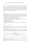
4 JOURNAL OF COSMETIC SCIENCE The first day, in order to obtain a gradient of energy for the MED determination, the black blotting paper with the six square holes was fixed on one forearm of the volunteer (randomized choice). Then each square was irradiated with an exposure time that varied with a geometric progression (1.25 rate) because the sun simulator used in this experi- ment delivered only one light beam. The MED for unprotected skin was read the second day of the test, 24 hours after UV exposure. Application. On the second day, two sites with dimensions of 2 x 2 cm were marked out on the other forearm. On the two sites, 2 pl/cm 2 of the product was applied with a finger glove, one hour before the beginning of exposure for one site and one hour before tape stripping for the other site (reference). Tape stripping. Fifteen tape strippings were performed one hour after product application on the non-irradiated site: a 2 x 1.9-cm piece of adhesive was cut with scissors and immediately applied on the skin. The tape was pressed for one minute with a 500-g mass, then stripped in one quick move with forceps and then put in a 10-ml amber flask. This was repeated on the same skin area 15 times, and the tapes were grouped by five, to be assayed from the 11th to the 15th tape, because the concentrations of UV filters in these tapes were supposed to be below the detection limit of HPLC. The finger glove was conserved in a 10-ml amber flask in order to be assayed as well. The other site was irradiated at 40 MED one hour after product application. Tape stripping was performed immediately after irradiation in the same manner as the non- irradiated site. Extraction. The organic filters were extracted by adding 5 ml of chloroform/methanol, in a ratio of 2:1 (v/v), to each flask, except for the flask containing the finger glove in which 9 ml was introduced in order to obtain a better extraction. The solutions were filtered on a 0.45-pm pore diameter filter in a 1-ml amber shell glass. HPLC method. The calibration was done with an external standard method using four standards. The programs for the mobile phase and the detector absorbency were determined in a preliminary study, and the reproducibility was found to be greater than 95%. Table II presents the final operating conditions entered in the Millenium program. General conditions: © Volume of injection: 5 pl © Temperature: 25øC © Wavelength of detection: 310 nm © Flow rate: 1.5/min Table II Operating Conditions of the Millenium Program (mobile phase) Time (rain) % of Methanol % of Water % of THF 0 80 20 0 3 80 20 0 5 80 0 20 10 80 0 20 12 80 20 0 15 80 20 0
Purchased for the exclusive use of nofirst nolast (unknown)
From: SCC Media Library & Resource Center (library.scconline.org)