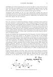
82 JOURNAL OF COSMETIC SCIENCE COSMETIC APPLICATIONS OF A WOUND HEALING PEPTIDE Karl Lintner, Ph.D., Philippe Mondon, Ph.D., Oliver Peschard, Ph.D. and Claire Mas-Chamberlain, Ph.D. Serderma, Le Perray en Yvelines, France Introduction Wound healing is a very complex process, the mechamcs of which have not yet been completely unravelled. Although some general features are common to the wound repair mechanisms in different organs and tissues, we shall focus here on the activities observed in skin tissue, which in any case, when we think of a "wound", comes to mind first. Wound healing process What happens when we cut our skin? The first step in damage control is to stop the potentially deadly loss of blood: coagulation occurs in the micro- vessels and fibrin is deposited to obdurate any holes or openings. This occurs through a cascade of protein activation steps. Peptides cleaved from circulating proteins during this process are not just by-products, but have their own purpose and activity. They are often chemotacfic, that is, they attract cells (platelets, leucoc3•tes, macrophages and fihroblasts) to the site of the wound. Some peptides then act on the damaged cells and provoke their death (apoptosis, a safety feature), others act on the remaining cells and on those chemically attracted cells to stimulate them into the synthesis of new tissue. Indeed, once the blood flow has been taken care of, inflammation (more or less pronounced depending on the size of the wound) sets in: Fibroblasts first secrete proteolytic enzymes to help clean out the wound, swelling due to increased cell density and blood flow occurs. After the damaged tissue has been removed and oedema has been circumscribed, the process of reconstruction of the conjuncfive tissue begins by deposition of collagen fibres and glycosaminoglycanes. The covering of the wound site by epithelial cells also initiates the synthesis of matrix macro-molecules in the fibroblasts • What makes the fibroblasts change their activity from "lytic" to "synthetic"? It appears that an extemal signal, like a local hormone, a messenger molecule that interacts with the fibroblast cell membrane receptors, induces this change. And what kind of signal might that be? Obvious candidates are small signal peptides, those that are released from the degradation process of the macromolecules. This feedback mechanism is a logical consequence of the wound-healing cascade and confirms that mother Nature is economical: use every waste product possible for some beneficial purpose! Some of these peptides, fragments of the degraded macromolecules, have now been isolated, identified and synthesised in larger quantities for further investigation. If we consider that the ageing process (induced by sun exposure and the ensuing inflammatory reactions, the free radicals, the upregulation of elastolysis and collagenolysis) is a slowly occurring wound (the "injuries" of time, Horace, Odes II1), then the use of these signalling "wound healing peptides" in cosmetics seems worth pursuing. We investigated the biological and cosmetic activity of a model peptide Ala-g-His (camosine) and three peptides that are fragments of elastin (Val-Gly-Val-Ala-Pro-Gly =VGVAPG, chemotactic), collagen I (central sequence: Gly-His-Lys =GHK) and procollagen I terminal sequence (Lys-Thr-Thr-Lys-Ser =KTTKS). For reasons of time and space we shall discuss only the latter one here. Material and Methods Peptides were synthesized by classical Merrifield solid phase methods. In vitro and ex vivo measurements of collagen and GAG synthesis are described elsewhere (2,3). Results and Discussion Skin diffusion In order to use biologically active peptides in cutaneous application at concentration levels that are safe, effective and nevertheless economical, optimum skin diffusion must be assured. For this purposes it is necessary to modify the peptide sequence in order to improve cutaneous substantivity and diffusion. We therefore added a palmitoyl chain to the N-terminal amino acid and then tested the in vitro, ex vivo and in vivo activity of the Pal- KTTKS molecule. In a previous model study we had compared the skin diffusion (using radiolabels) of a dipeptide Ala-His with its palmitoylated equivalent Pal-Ala-His.

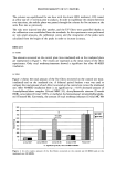
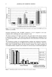
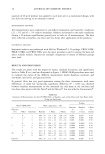
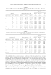
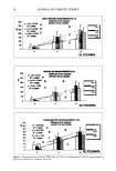
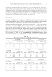
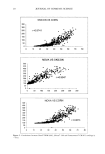
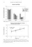
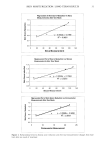
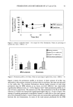
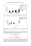
2000 ANNUAL SCIENTIFIC MEETING 83 We were able to show that diffusion into the skin is negligible with an unmodified peptide, whereas the palmitoylated peptide enters epidermis and dermis to significant extents and thus is able to deliver biological activity at physiological concentrations (2). Preliminary data on the Pal-KTrKS peptide confirm this observation. Collagen IV and GAG synthesis in vitro Pal-KTTKS was tested on normal human fibroblasts plated from a 63 year old skin biopsy. We were looking specifically for stimulation of the synthesis of Collagen IV, a major protein involved in the maintenance of epidermal-dermal junction. At the concentration levels of 104M/I (i.e. 2 to 4 ppm of peptide,) we observe a 2 to 4 fold increase in collagen IV synthesis as measured by immunoblot techniques. A 50% increase in GAG synthesis is also observed. In both studies, TGF 1• was used as positive control. Ex vivo Collagen I •nthesis In an ex vivo model (full thickness human skin biopsies of a 43 year old patient), strong collagen I synthesis is obtained at concentration levels of 2 and 4 ppm of Pal-KTTKS peptide in solution. Vitamin C at 1000 ppm increases collagen synthesis in this protocol by 42%, TGFg at 10 ppb by 33% 4 ppm of the Pal-KTTKS peptide achieve 117% increase in 3H-Proline incorporation, thus more than double the Vitarmn C activi,ty at 250 fold lower concentration. In vlvo studies Following thorough investigation of the non-toxicity and cutaneous tolerance of the palmitoylated peptide, three in vivo studies were carried out: ß one vehicle controlled half face study on 25 panellists, ß one half face study comparing the peptide containing cream against a 5% vitamin C cream on 10 panellists (both studies over 2, 4 and 6 months), and ß one 2 and 4 month study on 20 panellists comparing two identical base creams, one containing 3ppm of Pal-KTTKS peptide, the other containing 700 ppm of retinol. Parameters investigated by image analysis of skin replicas were wrinkle profile (depth, length, density, volume .... ), skin thickness and density as measured by 2D and 3D ultrasound echography, visual dermatological scoring and self assessment. All three studies concur: the peptide has skin tissue repair activi• of high potency. The first study showed that the vehicle cream (although moistunsing) showed no wrinkle reduction at all over 2, 4 or 6 months. Surprisingly, the commercial cream with Vitamin C did not improve the investigated skin parameters in any significant manner. The pepfide containing cream, however, leads to spectacular skin profile improvement in this time span that is evident from the study of skin replicas and from visual assessment by the dermatologist, the panellists and close-up photography. lIT 2 months mT 4 months Wrinkle Parameters Depth Length Volume Surface The third study, conducted a year later on a different panel and with a different vehicle cream confirmed the initial results. The peptide containing cream (3 ppm of Pal-KTTKS) leads to significant wrinkle reduction (figure 1), skin thickening and densification - in agreement with the expected collagen and GAG synthesis - over a period of 2 and 4 months. Visually detectable effects were observed by the attending dermatologist. The high dose of 700 ppm of retinol showed similar effects after 4 months and served as a positive control, as its effect on wrinkles is well known Conclusion Clearly the concept of using natural, biologically active peptides for cosmetic applications is a viable strategy leading to perceivable skin benefits. Specifically chosen peptides, acting as messenger molecules in normal biological processes such as growth and tissue repair are safe, natural, innovative molecules well adapted to the role of cosmetic active ingredients and thus an emerging alternative to unstable or (sometimes) overrated vitamins. [1] A. Sim6on et al. in: Current Topics in Pathology, Vol. 93, 95-101(1999), A. Desmouli•re et B. Tuchweber, eds. (•Springer Berlin Heidelberg [2] K. Linther and O. Peschard, Int. J. Cosrn. $ci. 22, 207-218, (2000) [3] P. Mondon et al. in preparation
Purchased for the exclusive use of nofirst nolast (unknown)
From: SCC Media Library & Resource Center (library.scconline.org)