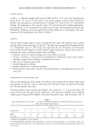
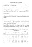
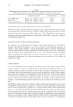
48 JOURNAL OF COSMETIC SCIENCE Table III Amount (pg) of tx-T as a Metabolite From tx-TAc in IPM Solution and as the Active From tx-T IPM solution Time (hr) Metabolite o•-T o•-T active 2 3.108 (+0.53) 2.468 (+0.43) 6 3.668 (+0.47) 1.612 (+0.40) 12 2.641 (+0.77) 3.317 (+0.60) 24 5.404 (+0.91) 2.543 (+0.30) Values are micrograms, mean _+ SEM (n = 4). membranes, enter cells, and be associated with other hydrophobic membranous struc- tures present in the cells such as the mitochondria and nuclear membranes. Hydrolysis may be catalyzed through intracellular esterases and/or lipases. At the end of 24 hours, however, there was no significant difference between the amount of active that permeated the viable skin from either the o•-TAc IPM solution (o•-TAc + o•-T in viable skin, 3.04 + 0.65) or o•-T IPM solution (o•-T in viable skin, 4.953 + 0.8). This is comprehensible if we take into account that the pH of the viable part of skin is about 7.4, and that the aromatic hydroxyl group in o•-T (pKa = 10) is not dissociated under these conditions. As o•-T is undissociated, the difference between o•-TAc and o•-T with regard to the physicochemical parameters, which determine transport, is negli- gible. Beijersbergen van Henegouwen et al. (25) found that o•-TAc and o•-T behaved similarly with regard to transport through the epidermis (i.e., penetration) and hori- zontal migration in the epidermis. In our results we also found that using a 1% o•-T formulation gives a higher concen- tration of o•-T in terms of micrograms of drug in viable skin than as a metabolite from a 5% o•-TAc formulation. Approximately 1.5% of o•-T yields the same viable skin concentration as 5% o•-TAc. This finding has important implications in the topical delivery of o•-T. To deliver larger amounts of o•-T in the skin, an o•-T formulation is more efficient than obtaining o•-T as a by-product of o•-TAc metabolism. However, this has to be weighed against stability problems with o•-T and the shelf life of the product. The most intriguing results of this study were that metabolism occurred as early as two hours on application of the ester and that the extent of bioconversion in relation to total skin concentrations reached a peak at about 6-12 hours. Longer time periods of up to 24 hours did not correspondingly increase the extent of metabolism. This could possibly be due to saturation of the metabolizing enzymes present in the skin, whose concen- trations are about two orders of magnitude lower than that in the liver. High prodrug concentration in skin may lead to enzyme saturation kinetics and, as a result, limited conversion of the o•-TAc into o•-T. Another possibility could be the waning effect of enzyme activity of the viable skin that may occur in in vitro studies at the end of 24 hours. This is a drawback of in vitro methods. In vivo, however, the extent of cutaneous metabolism is difficult to differentiate from systemic metabolism, and a better quanti- tative determination of cutaneous metabolism is still obtained in vitro. In a living skin equivalent model, maximum conversion of o•-TAc to o•-T was obtained at about six hours (15). Similar results were obtained by Kramer-Stickland and Liebier (26), who found maximum hydrolysis at three hours post-treatment and have suggested that hydrolysis in unirradiated mice may be a saturable phenomenon. They have alluded to

PERMEATION AND METABOLISM OF o•-T and o•-TAc 49 the possibility of the hydrolytic pathway being saturable even over a short period of 24 hours, and an increase in d3-o•-tocopheryl acetate concentration did not result in a corresponding increase in d3-o•-tocopheryl acetate hydrolysis. About 20-50% of the applied dose of the o•-TAc was found to be metabolized to o•-T when measured in terms of drug permeated in the stratum corneum and viable skin. This is about 2-5 fold higher than that found by Norkus eta/. (13) in hairless mouse skin. Tojo and Lee (27) studied the bioconversion of a provitamin to vitamins C and E in mouse skin dermis. The provitamin was inherently stable and was expected to develop actions of both vitamins E and C in the body through splitting off the phosphoric acid esters by enzyme phosphatase. They calculated the yield of bioconversion to be about 96% in the hairless mouse skin. In summary, this study has demonstrated the metabolism of o•-TAc to o•-T in viable pig skin. Topically applied o•-TAc was bioconverted to the active molecule and free radical scavenger o•-T within the skin tissue. No metabolism was detectable in the stratum corneum. This study has also elucidated the kinetics of metabolism of o•-TAc. The extent of metabolism was highest at 6-12 hours after application. Longer time periods failed to produce a higher extent of metabolism, probably due to the saturation of the hydro- lyric pathway. REFERENCES (1) A.L. Norins, Free radical formation in the skin following exposure to ultraviolet light, J. I,vest. DermatoL, 39, 445•448 (1962). (2) M.M. Marhews-Roth, Carotenoid pigment administration and the delay in development of UV-B- induced tinnors, Photochem. Photobio/., 42, 35-38 (1983). (3) N. Khettab, M.C. Amory, G. Brigand, B. Bousquet, A. Combre, A.Y. Forlot, and M. Barey, Photoprotective effect of vitamins A and E on polymamine and oxygenated free radical metabolism in hairless mouse epidermis, Biochimie, 70, 1709-1713 (1988). (4) E. Niki, "Function of Vitamin E as Antioxidant in the Membranes," in Vitami, E: Its Usef•/,es• Health a,d ], C•ri,g Diseases, M. Minoe, H. Nakamura, A. T. Diplock, and H. J. Kayden, Eds. (Karger, New York, 1992), pp. 23-30. (5) H. L. Gensler and M. Magdaleno, Topical vitamin E inhibition of immunosuppression and tumori- genesis induced by ultraviolet irradiation, N•tr. Ca,er, 15, 97-106 (1991). (6) D. L. Bissett, G. G. Hillerbrand, and D. P. Hannon, The hairless mouse as a model of skin photoaging: Its use to evaluate photoprotective materials, Photodermato/ogy, 6, 228-233 (1989). (7) P. Mayer, The effects of vitamin E on the skin, Cosmet. Toilerr., 108, 99-109 (1993). (8) M. Lopez-Torres, J. J. Thiele, Y. Shindo, D. Han, and L. Packer, Topical application of ½x-tocopherol modulates the antioxidant network and diminishes ultraviolet-induced oxidarive damage in murine skin, Br. J. Dermato/., 138, 207-215 (1998). (9) A. Parmatier, P. Jenner, B. Testa and J. C. Etter, The skin as a drug-metabolizing organ, Dr•g. Rev., 8, 319-343 (1978). (10) J. Rautio, H. Taipale, J. Gynther, J. Vepsalainen, T. Nevalainen, and T. Jarvinen, I, vitro evaluation of acyolxyalkyl esters as dermal prodrugs of ketoprofen and naproxen,J. Pharm. Sc]., 87, 1622-1628 (1998). (11) D. S. Alberts, R. Goldman, M-J. Xu, R. T. Dorr, J. Quinn, K. Welch, J. Guillen-Rodriguez, M. Aickin, Y-M. Peng, L. Loescher, and H. Gensler, Disposition and metabolism of topically adminis- tered ½x-tocopherol acetate: A common ingredient of commercially available sunscreens and cosmetics, N•tr. Ca,cer, 26, 193-201 (1996). (12) H.L. Gensler, M, Aicken, Y-M. Peng, and M. Xu, Importance of the form of topical vitamin E for prevention of photocarcinogenesis, N•tr. Ca,cer, 26, 183-191 (1996).
Purchased for the exclusive use of nofirst nolast (unknown)
From: SCC Media Library & Resource Center (library.scconline.org)