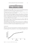
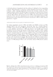
155 ANTIPIGMENTATION AND ANTIOXIDANT ACTIVITY Environmental exposure factors mainly include air pollution, ultraviolet (UV) irradiation, and multifarious physical and chemical stimulation These trigger many skin problems, such as skin hyperpigmentation, dryness and aging, and so forth (4). Pigmentation is a key factor in determining skin color, as well as one of the defense systems that protects skin from UV damage (5). Melanogenesis is a complex biological synthesis that includes melanocyte proliferation, melanin production, and melanosome transfer to keratinocytes. UV exposure can activate tyrosinase and promote melanin synthesis in melanocytes (6), leading to hyperpigmentation and skin disorders. The copper-containing enzyme tyrosinase is a key rate-limiting enzyme in melanin biosynthesis, which catalyzes the production of melanin through oxidation of L-tyrosine to L-dihydroxyphenylalanine (L-DOPA) and dopaquinone (7). Subsequently, L-DOPA is converted to either eumelanin or pheomelanin, depending on its reaction with cellular cysteine or dopachrome tautomerase and tyrosinase-related protein 1 (8). UV irradiation stimulates excessive reactive oxygen species (ROS) generation through alteration in cellular metabolism, which breaks down the cellular antioxidant defense system and triggers oxidative stress (9). DNA damage, lipid peroxidation, and apoptotic signal transduction are triggered, clarifying these thoughts owing to the oxidative imbalance and further influencing cellular physiological function (10). The accumulation of lipofuscin and melanin caused by superfluous ROS can turn into senile plaques, melasma, dark spots, and other pigmented skin diseases (11), eventually leading to skin photoaging and skin cancer. However, skin cells can upregulate cellular antioxidant capacity through glutathione peroxidase (GSH-Px), total antioxidant capacity (T-AOC), superoxide dismutase, catalase, and nonenzymatic mechanisms in order to maintain a normal physiological environment (12). The authors evaluated the inhibitory ability of SFE for melanin production at the cellular level and even in the reconstructed melanin skin model. Its antioxidant capacity was also assessed. The double effects of SFE indicated that it had the potential to become an effective cosmetic ingredient. MATERIALS AND METHODS MATERIALS Dried SFE was collected from Yunnan Baiyao Group Health Products Co., Ltd (Kunming, Yunnan, China). Dulbecco’s Modified Eagle Medium (DMEM) (Corning®, Corning, New York), penicillin, streptomycin, and fetal bovine serum (FBS) were purchased from Gibco (Waltham, Massachusetts). Other chemicals were from Servicebio (Hubei, China). MelaKutis® (Biocell Biotechnology, Guangdong, China) was manufactured by Biocell Biotechnology (Dongguan, Guangdong, China). CELL VIABILITY Cytotoxicity of SFE to B16-F0 and HaCaT cells. B16-F0 cells were seeded at 2 × 10⁴ cells per well, and HaCaT cells were placed at 2 × 10⁵ cells per well in DMEM complete medium in 96-well plates. They were cultured in a humidified environment at 37°C and 5% CO 2 for 24 hours and then treated with SFE in serum-free DMEM for 48 hours (13). Cell viability was measured by the cell counting kit (CCK)-8 assay. Both B16-F0 and HaCaT cells were incubated with 10 µL CCK-8 solution per well for 2.5 hours, and absorbance was measured at 450 nm by the Thermo Fisher Scientific (Waltham, Massachusetts) microplate reader.

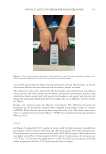
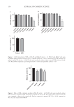
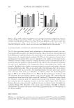
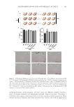
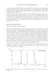
156 JOURNAL OF COSMETIC SCIENCE Cytotoxicity of UVB exposure to HaCaT cells. HaCaT cells were placed at 2 × 10⁵ cells per well in DMEM complete medium in 96-well plates at 37°C with 5% CO 2 for 24 hours. The cells were then rinsed twice with phosphate buffer saline (PBS) and covered with a thin layer of that PBS. A UV radiometer (Z03-II, SCIENT) was used for UVB irradiation (50 mJ/cm2). Afterward, HaCaT cells were incubated with 10 µL CCK-8 solution per well for 2.5 hours, and absorbance was measured at 450 nm by the Thermo Fisher Scientific microplate reader. MELANIN CONTENT B16-F0 cells were seeded at a density of 2 × 10⁴ cells per well in 6-well plates and incubated for 24 hours in DMEM (10% FBS and 1% penicillin streptomycin) (14). The cells were cultured with SFE at different concentrations or positive control (PC) arbutin in serum-free DMEM for 48 hours. After treatment, the cells were collected and washed twice with PBS. To assess the melanin content, the cells were dissolved in lysis buffer (1M NaOH, 10% DMSO) at 80°C for 30 minutes. Absorbance was measured at 405 nm. CELLULAR TYROSINASE ACTIVITY B16-F0 cells were seeded at a density of 2 × 10⁴ cells per well in a 6-well plate and incubated overnight in DMEM (10% FBS and 1% penicillin streptomycin) (15). The cells were incubated with SFE or PC arbutin in serum-free DMEM for 48 hours. They were then isolated with trypsin, washed with PBS twice, and lysed with 1% Triton X-100® (Dow Chemical Company, Midland, Michigan). Then, 100 µL of each lysate (preheat to 37°C for 5 minutes) was mixed with 100 µL of 0.25% L-DOPA in a 96-well plate and incubated at 37°C for 1 hour. Absorbance was measured at 490 nm. ANTIOXIDANT ENZYMES ACTIVITIES The HaCaT cells were placed in 6-well plates at a density of 2 × 10⁵ cells/well with DMEM (10% FBS and 1% penicillin streptomycin) for 24 hours (16). The cells were incubated with serum-free medium, including SFE or PC ascorbic acid in serum-free DMEM for 6 hours. Then, they were rinsed twice with PBS and exposed to UVB radiation (50 mJ/cm2). After UVB exposure, cells were homogenized in cold lysis buffer. The GSH-Px and T-AOC activities were detected by ELISA kit according to the manufacturer’s instructions. EFFECT OF SFE IN THREE-DIMENSIONAL SKIN MODEL (MELAKUTIS) To investigate the effects of SFE on melanin synthesis and deposition, the reconstructed human skin model of MelaKutis (17) was incubated with SFE or kojic acid, respectively. MelaKutis was cultured in M-TA medium (Biocell Biotechnology, China) at 37°C in 5% CO 2 . Then, 10 µL of different concentrations of SFE (0.5, 1, or 2 mg/mL) or PC kojic acid (500 µg/mL) were added on the surface of each model. Three-dimensional (3D) skin models were irradiated by UVB radiation (50 mJ/cm2), and the media were changed every day for 6 days. SFE (0.5, 1, or 2 mg/mL) or kojic acid was added at day 3 and day 5. On day 7, all
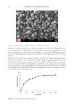
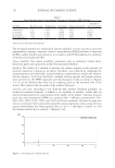
Purchased for the exclusive use of nofirst nolast (unknown)
From: SCC Media Library & Resource Center (library.scconline.org)