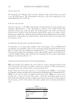
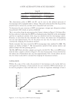
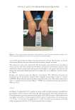
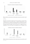
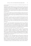
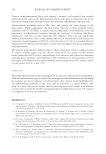
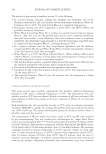
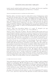
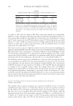
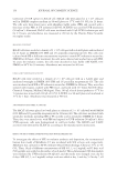
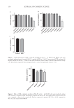
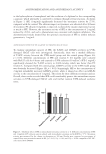
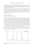
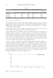
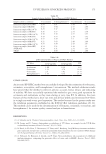
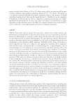
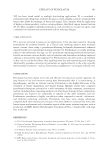
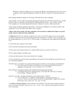
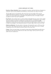
157 ANTIPIGMENTATION AND ANTIOXIDANT ACTIVITY skin models were collected and subjected to appearance observation, brightness, melanin content, and melanin distribution. Melanin content. The collected models were dissolved in 1M NaOH containing 10% DMSO at 80°C for 40 minutes. The absorbance of supernatant was measured at 405 nm. Measurement of skin color. The pigmentation intensities in 3D skin models were measured by a colorimeter (DSM II, Denmark) and were expressed as the L* values. Fontana–Masson staining. The 3D skin models were fixed with 10% buffered formalin and embedded in paraffin. Melanin was visualized using Fontana–Masson staining with an eosin counterstain according to the manufacturer’s instructions. STATISTICAL ANALYSIS Statistically significant differences between two groups were determined by a student’s t-test, where p 0.05 was considered statistically significant. All analyses were performed using GraphPad Prism 9.0 (GraphPad Software LLC, San Diego), with the experimental results expressed as mean ± SD (n ≥ 3). RESULTS CELL VIABILITY B16-F0 melanoma cells were used to evaluate the inhibitory effect of SFE on melanogenesis, and HaCaT cells were applied to access the antioxidant activity of SFE. The cytotoxicity of either the medicine or cosmetic agent is of chief importance when used. The effects of SFE on the viability of B16-F0 and HaCaT cells were determined by CCK-8 assay. As shown in Figure 1A-B, SFE had no significant cytotoxic effect on B16-F0 and HaCaT cells at the concentrations from 0.0625 to 1 mg/mL. SFE at 2 mg/mL was slightly toxic to B16-F0 and HaCaT cells, but the toxicity was within acceptable limits. UV irradiation (50 mJ/cm2) did not cause obvious damage to HaCaT cells under this condition, as shown in Figure 1C. ANTIMELANOGENESIS EFFECT OF SFE IN B16-F0 CELLS Murine B16-F0 melanoma cells were used to investigate the inhibitory effect of SFE on melanogenesis. Treatment with SFE (1 mg/mL) for 48 hours significantly decreased the melanin content of B16-F0 cells by 25.5%, compared with the control group without cytotoxicity, while PC arbutin (1 mg/mL) reduced the melanin content by 23.3% (Figure 2). Unexpectedly, the reduction of melanin content of SFE (2 mg/mL) only reached 9.9%, which was less effective than SFE at a low concentration. These results indicated that SFE (1 mg/ mL) was relatively safe and more effective than PC arbutin in inhibiting melanin production. ANTITYROSINASE ACTIVITY OF SFE IN B16-F0 CELLS To clarify the mechanism underlying the observed SFE-induced depigmentation, the expression of melanogenesis-related tyrosinase was determined. This enzyme is involved

158 JOURNAL OF COSMETIC SCIENCE Contro l 0.0625 mg /m L 0.12 5 mg / mg/m L mg/m L 1 mg2 / mg/m L 4 m g/mL 0 50 100 Co n t l 0. 625 m g/mLmg/m 0. 1 2 L 0.25 m g/mL 0 mg/m L 1 m g/mLg/mL 2 m mg/m L 0 50 100 a b Control UVB 0 50 100 c Figure 1. (A,B) Cytotoxicity of SFE on B16-F0 and HaCaT cells (n = 5). B16-F0 and HaCaT cells were incubated without (control) or with 0.0625–4 mg/mL of SFE for 24 h. (C) Cytotoxicity of UVB radiation (50 mJ/cm2) on HaCaT cells (n = 3). HaCaT cells were irradiated with or without (control) UVB radiation (50 mJ/ cm2). Results were expressed as percentages relative to control and presented as mean ± SD. Negati ve controArbuti2 l n mg /m L 1 mg/m L 0 50 100 *** *** *** Figure 2. Effect of SFE on melanin synthesis in B16-F0 cells (n = 4). B16-F0 cells were incubated without (NC) or with 1–2 mg/mL of SFE, and arbutin 1 mg/mL was used as PC. Percentage values in the treated cells were compared to those in the control cells. Data are expressed as mean ± SD ***p 0.001 compared to NC. NC: cells treated with DMEM. B16 Cell viability% HaCaT Cell viability% HaCaT cell viability % Melanin content %
Purchased for the exclusive use of nofirst nolast (unknown)
From: SCC Media Library & Resource Center (library.scconline.org)