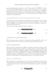
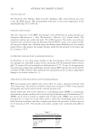
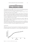
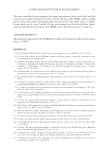
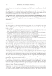
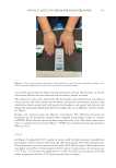
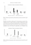
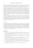
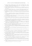
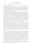
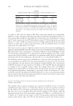
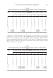
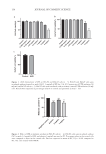
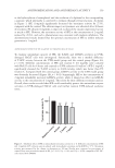
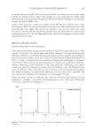
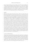
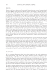
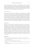
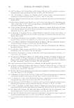
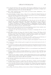
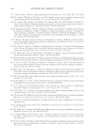
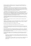
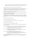
159 ANTIPIGMENTATION AND ANTIOXIDANT ACTIVITY in the hydroxylation of monophenol and the oxidation of o-diphenol to the corresponding o-quinone, which ultimately is converted to melanin through several reactions. As shown in Figure 3, SFE (1 mg/mL) significantly decreased the tyrosinase activity by 27.3%, compared with the control. The obvious impact on tyrosinase was observed after 48 hours of treatment. PC arbutin (1 mg/mL), as expected, weakened the enzyme expression, but not as much as SFE. However, the tyrosinase activity of SFE at the concentration of 2 mg/mL reduced by 20.6%, and such a phenomenon was consistent with melanin inhibition. The aforementioned results showed that the optimal concentration of SFE to inhibit melanin generation is 1 mg/mL. ANTIOXIDANT EFFECT OF SFE AGAINST UV-TREATED HACAT CELLS To examine antioxidant capacity of SFE, the T-AOC and GSH-Px activities in UVB- damaged HaCaT cells were investigated. Statistically, there was a marked difference of T-AOC activity between the UVB model group and the control group (Figure 4A, p 0:05). Different concentrations of SFE and vitamin C (0.1 mg/mL) were cultured with HaCaT cells for 6 hours and exposed to UVB radiation (50 mJ/cm2). SFE (1 mg/mL) significantly elevated the T-AOC activity to 0.060 mmol/g, which was better than PC vitamin C. Compared with the control group, GSH-Px activity in the UVB model group was obviously decreased (Figure 4B, p 0.01). Surprisingly, SFE at the concentration of 2 mg/mL remarkably increased GSH-Px activity, while it showed no effect on GSH-Px activity at the concentration of 1 mg/mL. The reason for these differences remains unclear. Overall, these results revealed that SFE could considerably protect the antioxidant enzyme activities in UVB-damaged HaCaT cells and further restrain UVB-induced oxidative stress. Negati ve controArbu l ti 2 mg/m1 L mg/m L 0 50 100 *** ** * Figure 3. Inhibitory effect of SFE on intracellular tyrosinase activity (n = 4). Different concentrations of SFE and 1 mg/mL of PC arbutin were incubated with intracellular tyrosinase and L-DOPA at 37°C. Tyrosinase activity was measured by the change in absorption at 490 nm. Results were expressed as percentages of control. Data were presented as mean ± SD *p 0.05, **p 0.01, and ***p 0.001 compared to NC. NC: cells treated with DMEM. Tyrosinase content %

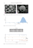
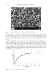
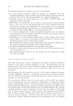
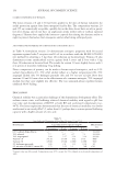
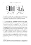
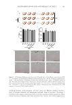
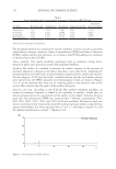
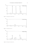
160 JOURNAL OF COSMETIC SCIENCE 3D SKIN EQUIVALENT TO INVESTIGATE THE WHITENING EFFICACY OF SFE The 3D skin equivalents showed many advantages in dermatological research over two- dimensional (2D) monolayer cell cultures for bioassay. This is because of their stratified and well-differentiated epidermal barrier (18), which reflects the morphological and molecular characteristics of a normal human epidermis (19). The use of reconstructed tissues also is advantageous in situations where an active ingredient is not soluble in cell culture media. Therefore, it may be soluble in an oil or a complex base that can be accommodated easily by a reconstructed tissue platform compared with the cell cultures. This provides the possibility for more active ingredients to be evaluated for efficacy and broadens the cosmetic raw materials. MelaKutis has a highly consistent histological structure and melanin response function with human skin. This can accurately reflect the whitening effect of the extract on the human body and can realize the multidimensional detection (gene level, protein level, cell level, tissue level, etc.). Therefore, we further evaluate the antimelanogenesis effect of SFE in the 3D skin model in vitro. When 3D skin models were irradiated with UV radiation, the total melanin content in the negative control (NC) group obviously increased more than that in the blank control (BC) (without UVB exposure) group (Figure 5C). However, treatment with SFE significantly decreased the melanin content, which was consistent with the brightness variation in the skin model (Figure 5A-B). Moreover, Fontana–Masson staining revealed a significant decrease in melain content throughout the epidermis (Figure 5D), efficiently in the cuticle, upon treatment with SFE at different concentrations. In general, the extract could reduce pigmentation due to UVB exposure in the 3D skin models. DISCUSSION Melanin, as a dark pigment, provides protection against harmful UV irradiation and DNA damage (20). However, abnormal accumulation of melanin causes dermatological problems Negati ve contro l UV B Vitamin C 2 mg/m L 1 mg/m L 0.00 0.02 0.04 0.06 # * * UVB Negati ve c o n t l UVB Vi t in C 2 mg/m L 1 mg/m L 0 2000 4000 6000 ## ** * UVB a b Figure 4. Effects of SFE on T-AOC and GSH-Px activities in UVB radiation-induced HaCaT cells. Cells were treated with different concentrations of SFE (2 and 1 mg/mL) or PC vitamin C (0.1 mg/mL) for 24 h and then irradiated with UVB radiation (50 mJ/cm2). (A) T-AOC activity and (B) GSH-Px activity and were tested. Data were shown as the mean ± SD (n = 3) ## p 0.01 and # p 0.05 versus the control **p 0.01 and *p 0.05 versus the UVB group. As an NC, HaCaT cells were treated with DMEM without UVB irradiation. T-AOC/(mmol/ GSH-Px/(U/mg protein)
Purchased for the exclusive use of nofirst nolast (unknown)
From: SCC Media Library & Resource Center (library.scconline.org)