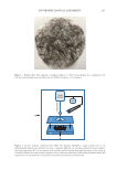
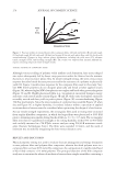
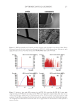
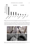
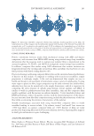
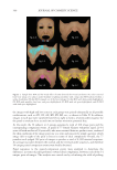
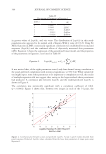
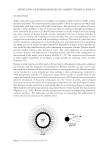
317 Microsponge Loaded Topical Gel METHODS OF PREPARATION OF BENZOYL PEROXIDE–LOADED MICROSPONGES Benzoyl peroxide microsponges were prepared by dissolving the drug (2.5 g) in polymer PLGA (1 g), which was previously dissolved in the methanol. The resulting solution was added to the aqueous phase containing 0.1 mL of span 80 and 1 g of 1% w/v PVA as an emulsifying agent. The mixture was then agitated using a propeller with a rotation speed of 500 rpm and 1,000 rpm (16). The dispersed drug and the polymer were immediately transformed into fine droplets, which subsequently solidified into rigid microsponges. Due to solvent evaporation, the particles were collected by filtration and washed with demineralized water and desiccated at room temperature for 24 hours (17). CHARACTERIZATION STUDIES OF MICROSPONGES: ENTRAPMENT EFFICIENCY Entrapment efficiency purified form of prepared vesicular suspension was used for the evaluation of percent drug entrapment. Vesicular suspension was centrifuged at 4,000 rpm for 1 hour. The obtained sediment after centrifugation was then lysed with the help of methanol. The solution thus obtained was filtered using a nylon filter disc (0.22 µm) and analyzed (18). Entrapment efficiency was found using the following equation: %Entrapment efficiency Wa (Ws Wp)/Wa], =-+[where Wa =total drug added Ws =drug present in supernatant and Wp =drug present in sediment. IN VITRO DRUG RELEASE STUDIES FOR MICROSPONGES The dialysis membrane was used in Franz diffusion cell (19). The formulation containing benzoyl- loaded PLGA (microsponges) was applied on the dialysis membrane and then fixed in between the donor and receptor compartment of the diffusion cell. The receptor compartment contained a phosphate buffer (100ml) of pH 5.5. The diffusion medium temperature was thermostatically controlled at 37° ± 1° by surrounding water in jacket, and the medium was stirred at 500 rpm by a magnetic stirrer (20). The samples were withdrawn at predetermined intervals and replaced by an equal volume of fresh fluid. The samples withdrawn were spectrophotometrically estimated at 235 nm against their respective blank (21). SCANNING ELECTRON MICROSCOPE The surface morphology of microspheres was investigated using a scanning electron microscope (SEM). To prepare specimens for the polarizing, the microsponge was first taken on the slide as powder form and placed on the base plate. A vacuum was created through the system to reduce the conduction. Images were created scanned at different magnifications (22).

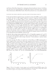
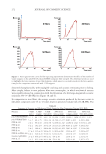
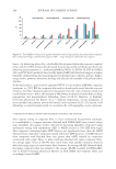
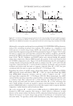
318 JOURNAL OF COSMETIC SCIENCE DRUG CONTENT STUDY A drug content study was done to determine the amount of the drug present in certain quantities of the formulation. One g of the formulation was taken into a 10 mL volumetric flask to that methanol was added, shaken well, and the volume was made up with the required amount of methanol. The volumetric flask was kept for 2 hours and shaken well in a shaker to mix it properly (23). The solution was passed through the filter paper and then filtered. Finally the mixer was measured for its absorbance by using a spectrophotometer at 235 nm (12). DRUG CONTENT SAMPLE ABSORBANCE STANDARD ABSORBANCE =×100 VALIDATION OF EXPERIMENTAL DESIGN Using Design Expert software version 11 (SRIHER, Chennai, India), the polynomial equation was generated for a dependent variable. A statistical model comprising of interactive and polynomial terms was used to evaluate the responses for the experiment. As a result, polynomial and transformed polynomial equations were generated. Extra design check point formulations were developed to validate the obtained polynomial equation model. Developed formulations were evaluated for dependent variable (i.e., percent entrapment efficiency and in vitro drug release (24)). The values obtained were then compared with the predicted value obtained from the transformed polynomial equation and evaluated statistically by analysis of variance (ANOVA 25). SELECTION AND CHARACTERIZATION OF OPTIMIZED FORMULATION Optimized formulation was selected on the basis of desirability obtained through the experimental design. It was then subjected to validation statistically. Formulation exhibiting the highest desirability was selected as an optimized formulation (Run 1) and subjected to characterization for surface characterization, stability, and preformulation studies. Optimized formulations were characterized using an SEM, in vitro drug release, and entrapment efficiency (26). For surface morphological analysis, an SEM was used to prepare specimens for polarizing the microsponge was first taken on the slide in powder form and then placed on the base plate. A vacuum was created through the system to reduce the conduction. Images were created scanned at different magnification. Microscopic examination was done using a light microscope at magnification 100×. They were further evaluated for in vitro drug release and percent entrapment efficiency. The selected optimized formulation was then incorporated into a suitable carbopol gel (27). PREPARATION OF GEL CONTAINING DRUG-LOADED MICROSPONGES Purified water (60 mL) was heated to 75–85°C and methylparaben, propylparaben, and disodium edetate were dissolved into it. Carbopol 934 (1 g) was then added and dispersed
Purchased for the exclusive use of nofirst nolast (unknown)
From: SCC Media Library & Resource Center (library.scconline.org)