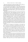
192 JOURNAL OF THE SOCIETY OF COSMETIC CHEMISTS (11) Herrmann, F., Prose, P. H., and Sulzberger, M. B.,Ibid., 21,397 (1953) (12) Harber, L. C., Herrmann, F., Mandol, L., and Sulzberger, M. B., Ibid., •9, 55 (1957). (13) Herrmann, F., Harber, L. C., Scher, R., Mandol, L.,/1. M./1./lrch. Derrnatol., 76, 282 (1957). (14) Wolf, J., Z. rnikroskop. anat. Forsch., 47, 351 (1940). (15) Szakall, A., Fette u. Seifen, 53, 399 (1951). (16) Pinkus, H., 2 t. Invest. Derrnatol., 16, 383 (1951). (17) Buckup, H., and Szakall, A., Berufsderrnatosen, 4, 1 (1956). (18) Herrmann, F., .drch. DerrnatoL SyphiloL, 200, 3 (1955). (19) Nicolaides, N., and Wells, G. C., •7. Invest. Derrnatol., 29, 423 (1957). (20) Montagna, W., "The Structure and Function of the Skin," New York, Academic Press, Inc. (1956). (21) Steigleder, G. K., and L6fFler, H., ,4rch. klin. u. exptl. DerrnatoL, 203, 41 (1956). (22) Steigleder, G. K., •7. Invest. Derrnatol., $1,29 (1958). (23) Kooyman, D. J., ./1. M. ./1. .drch. Derrnatol. SyphiloL, 25, 444 (1932). (24) Rothman, S., "Physiology and Biochemistry of the Skin," Chicago, University of Chi- cago Press (1954). RESPONSE OF THE HUMAN SEBACEOUS GLAND TO EXPERIMENTAL STRESS By JOHN S. STRAUSS, M.D.* Presented October 8, 1958, Seminar, New York City THE PURPOSE of this report is to detail the specific pathologic re- action patterns of the sebaceous glands following various experimental stresses. Furthermore, the responses will be correlated into a concise concept of response of the sebaceous gland after injury. However, before this is done, a few facts about the embryology, structure and function of the sebaceous glands must be reviewed. In embryonic life localized increased mitotic activity of the basal cell layer results in a bulging of the epidermal cells into the primitive derreal tissue. This is the primary epithelial germ which then grows down as a solid cord of basal cells. Part of the cord differentiates into a hair, other parts of this epithelial column form the sebaceous glands. Thus in embryonic life the entire pilosebaceous apparatus, including the hair and sebaceous gland arise directly from the epidermis. While the sebaceous glands vary greatly in size from area to area, the glands of the face and scalp are among the largest found in the body. Those in the scalp are associated with large terminal hairs, but on the glabrous portions of the face, particularly the cheek, the sebaceous glands reach enormous proportions and are the dominant structures in the follicles. Here the pilosebaceous duct leads * Dept. of Dermatology, Boston University School of Medicine--Massachusetts Memorial Hospitals, Boston 18, Mass. This study was supported under Grant E-1936C1 from the National Institutes of Health, United States Public Health Service.

192 JOURNAL OF THE SOCIETY OF COSMETIC CHEMISTS (11) Herrmann, F., Prose, P. H., and Sulzberger, M. B.,Ibid., 21,397 (1953) (12) Harber, L. C., Herrmann, F., Mandol, L., and Sulzberger, M. B., Ibid., •9, 55 (1957). (13) Herrmann, F., Harber, L. C., Scher, R., Mandol, L.,/1. M./1./lrch. Derrnatol., 76, 282 (1957). (14) Wolf, J., Z. rnikroskop. anat. Forsch., 47, 351 (1940). (15) Szakall, A., Fette u. Seifen, 53, 399 (1951). (16) Pinkus, H., 2 t. Invest. Derrnatol., 16, 383 (1951). (17) Buckup, H., and Szakall, A., Berufsderrnatosen, 4, 1 (1956). (18) Herrmann, F., .drch. DerrnatoL SyphiloL, 200, 3 (1955). (19) Nicolaides, N., and Wells, G. C., •7. Invest. Derrnatol., 29, 423 (1957). (20) Montagna, W., "The Structure and Function of the Skin," New York, Academic Press, Inc. (1956). (21) Steigleder, G. K., and L6fFler, H., ,4rch. klin. u. exptl. DerrnatoL, 203, 41 (1956). (22) Steigleder, G. K., •7. Invest. Derrnatol., $1,29 (1958). (23) Kooyman, D. J., ./1. M. ./1. .drch. Derrnatol. SyphiloL, 25, 444 (1932). (24) Rothman, S., "Physiology and Biochemistry of the Skin," Chicago, University of Chi- cago Press (1954). RESPONSE OF THE HUMAN SEBACEOUS GLAND TO EXPERIMENTAL STRESS By JOHN S. STRAUSS, M.D.* Presented October 8, 1958, Seminar, New York City THE PURPOSE of this report is to detail the specific pathologic re- action patterns of the sebaceous glands following various experimental stresses. Furthermore, the responses will be correlated into a concise concept of response of the sebaceous gland after injury. However, before this is done, a few facts about the embryology, structure and function of the sebaceous glands must be reviewed. In embryonic life localized increased mitotic activity of the basal cell layer results in a bulging of the epidermal cells into the primitive derreal tissue. This is the primary epithelial germ which then grows down as a solid cord of basal cells. Part of the cord differentiates into a hair, other parts of this epithelial column form the sebaceous glands. Thus in embryonic life the entire pilosebaceous apparatus, including the hair and sebaceous gland arise directly from the epidermis. While the sebaceous glands vary greatly in size from area to area, the glands of the face and scalp are among the largest found in the body. Those in the scalp are associated with large terminal hairs, but on the glabrous portions of the face, particularly the cheek, the sebaceous glands reach enormous proportions and are the dominant structures in the follicles. Here the pilosebaceous duct leads * Dept. of Dermatology, Boston University School of Medicine--Massachusetts Memorial Hospitals, Boston 18, Mass. This study was supported under Grant E-1936C1 from the National Institutes of Health, United States Public Health Service.
Purchased for the exclusive use of nofirst nolast (unknown)
From: SCC Media Library & Resource Center (library.scconline.org)