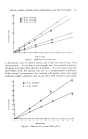
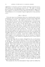
THERAPEUTIC POTENTIALITIES OF TRIGLYCERIDES 309 triglycerides, since they are inhibited by the fungistatic fatty acids, and again a simple laboratory test showed this to be true. We then had formulations or dosage forms of glycerol triacetate made in the University of Wisconsin Department of Pharmacy and submitted them to Dr. Sture A.M. Johnson of the University Hospitals Department of Dermatology for clinical testing against typical infections by the superficial dermato- phytes. The outcome of these early tests was very gratifying indeed the percentage of cures as judged by freedom from symptoms and negative microscope examinations of scrapings was about 90 per cent and there was no irritation or sensitivity. EXPERIMENTAL The dermatophytes used in this work were all fungi imperfecti and all are capable of producing specific "ringworm" infections or tineas. The common dermatophytes were obtained from the University Department of Dermatology, the others from the Communicable Disease Center at Cham- blee, Georgia. The cultures were maintained and tested on Sabouraud's medium. Table I shows the degree to which these fungi are inhibited when tri- acetin in varying amounts is incorporated into Sabouraud's medium. T•,BLE 1.--THE INHIBITION OF FUNGI BY TRIACETIN IN SABOURAUD'S DEXTROSE AGAR Diameter of Colony in mm. Triacetin, Per Cent Fungus 0 0.05 0.1 0.25 0.5 Epidermophyton floccosum 57 40 19 3 0 Trichophyton mentagrophytes 84 81 66 12 0 Trichophyton rubrum 71 73 59 9 0 Trichophyton tonsurans 34 29 17 5 0 Trichophyton verrucosum 21 13 11 0 0 Microsporum audouini 80 71 52 7 0 Microsporum canis 79 73 37 14 0 Microsporum gypseum 74 70 47 20 3 Candida albicans 20 22 20 16 9 It is rather difficult to compare the response of different fungi to triacetin because they grow at varying rates and the measurements of colony diam- eter were made at different times. There was considerable variation in the response to triacetin, but in general inhibition became apparent at about 0.1 per cent. There was also no obvious relationship between the inhibition and the type of infection typically produced by the various fungi. The effect of triacetin is upon the vegetative or growing cells the spores of Trichophyton mentagrophytes and Microsporum audouini have been soaked in triacetin for twenty-four hours and •were able to germinate when washed and placed on nutrient medium. Two independent attempts have been made to induce certain dermatophytes and Candida a/bicans to grow in

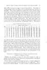
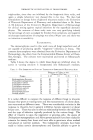
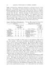
310 JOURNAL OF THE SOCIETY OF COSMETIC CHEMISTS higher concentrations of glycerol triacetate or to become resistant to the glyceride. All attempts to induce resistance of the fungi to 0.1 per cent glycerol triacetate have failed in fact, often the first colony on medium containing the glyceride is about 10 per cent larger than subsequent colonies derived from the first. Likewise attempts to induce resistance by the gradient plate procedure have been negative. It is apparent t•om these tests that triacetin certainly is not in the antibiotic class in regard to its effective concentration in laboratory tests but clinically results have been most gratifying. Here again, as many have noted in the past, there seems to be no constant relationship between in vitro and in vivo activity. TABLE 2.--TI•E EFFECT OF BLOOD SERUM ON THE FUNGISTATIC ACTIVITY OF 0.1 PER C•NT TRIACETIN Fungus TABLE 3.--MILLILITERS N/10 ACETIC ACXD PRODUCED BY ESTERASES FROM DERMATOPHYTES AND SERUM Diameter of Colony N/10 Acetic in mm. Acid Serum Added, Released in •----Per Cent------- ml. 0 1 5 10 Source of Esterase pH 6.5 pH 4.0 Epidermophyton Microsporum audouini 15.6 O . 4 floccosum 22 8 0 0 Trichophyton mentagrophytes 7.3 3.0 Trichophyton Trichophyton rubrum 6 8 O. 7 mentagrophytes 70 63 47 32 Serum, 5% 4.9 5.6 Microsporum Medium t¾om Microsporum audouini 49 21 18 13 audouini culture 12.2 0.2 Candida albicans 19 24 17 14 Boiled serum and fungi (control) 0 0 'Fable 2 shows the effect of blood serum on the fungistatic effect of 0.1 per cent triacetin. The extent of inhibition obtained in the presence of serum is the net result of opposing forces: the stimulatory effect of serum components on the fungus, the inhibitory effect caused by the increased acetic acid released by serum esterase and the inactivation, if any, of the serum on either acetic acid or triacetin. It is apparent that the inhibition caused by the increased acetic acid released by serum esterase was the dominant force. When triacetin and aliquots of serum were incubated together for twenty-four hours before addition to the test medium the results were the same, indicating that serum probably does not bind or otherwise make the triglycefide unavailable for hydrolysis. Table 3 shows that at least some of the superficial dermatophytes as well as blood serum contain esterase capable of releasing acetic acid from tri- acetin. Here again the fungi should not be compared, because no attempt was made to use the same number of cells or mass of tissue in each test. It is interesting to note that this strain of 3Iicrosporum audouini excretes considerable esterase into the medium in which it is growing if this were to occur in the infection also, the breakdown of epidermal lipids might play a part in the overall infectious process. It is noteworthy also that Candida
Purchased for the exclusive use of nofirst nolast (unknown)
From: SCC Media Library & Resource Center (library.scconline.org)