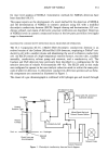
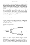
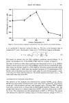
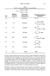
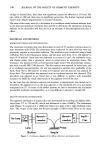
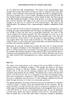
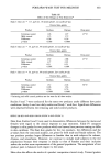
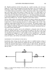
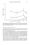
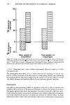
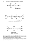
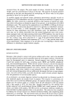
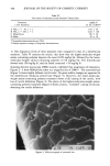
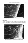
348 JOURNAL OF THE SOCIETY OF COSMETIC CHEMISTS vitiligo to normal skin, that there was significant protection afforded at 254 and 366 nm, while at 300 nm there was no significant protection. He further reported similar results with delayed pigmentation in normal volunteers. The aims of this study were (a) to determine if a correlation exists between melanin level in the skin and minimum erythema dose and (b) to determine the absorption of human melanin in the ultraviolet and thus arrive at an estimate of the photoprotection that it offers. MATERIALS AND METHODS MINIMUM ERYTHEMA DOSE DETERMINATION The minimum erythema dose was determined on each of 47 psoriatic patients about to start treatment with UVB. The phototests were conducted on skin sites that were not previously exposed to ultraviolet radiation. The irradiation was conducted using a bank of Philips TL12 4-foot fluorescent lamps, and the doses were from 10 to 150 mJ/cm 2 in steps of 10 mJ/cm 2. Each dose was delivered to an area of ! cm 2. An arithmetic series was chosen rather than a geometric series to avoid errors in irradiation times. The irradiance was measured with an International Light model 442 phototherapy radiom- eter with a model SEE 1240 detector. The skin reaction was assessed 24 hours later, and if no erythema was perceptible, the test was repeated in another area, preferably of the back, from !40 to 290 mJ/cm 2 in steps of 10 mJ/cm 2 and the reaction was assessed 24 hours later. This procedure was repeated until an erythema reaction was observed. This procedure was adopted as we found that it was difficult to predict with reasonable accuracy the MED by visual observation of the pigment level of the skin. At the time that the erythema reaction was assessed, a measurement was carried out to determine the pigment level in the skin adjacent to the irradiated area on a previously unexposed site (7). In some of the darker patients we had to determine the minimum perceptible erythema instrumentally, as it was difficult to discern it visually (8). MELANIN SPECTRUM DETERMINATION The apparent absorbance of human skin was determined by recording the diffuse reflec- tance from 275 to 720 nm (8), which was referenced to that of BaSO 4. The instrument used (Figure !) consisted of a !000-watt Xenon arc lamp with a light feedback stabi- lizer. The output of the lamp was focused through a 0.08-m water filter on the input Xe-Lamp ...m ....... •. Pr.o6• S•:mple Fief. Figure 1. A schematic diagram of the diffuse reflectance spectrophotometer.

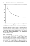
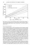
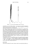
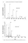
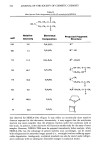
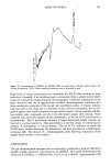
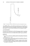
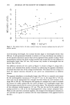
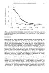
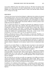
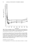
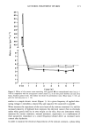
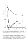
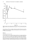
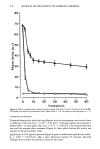
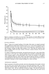
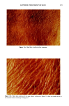
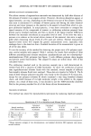
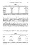
PHOTOPROTECTION BY HUMAN MELANIN 349 slit of a Jobin-Yvon HL monochromator. The output of the monochromator went through order-sorting filters before entering one end of a bifurcated fused silica fiber bundle, 3 mm in diameter. The joined end of the fiber was held in a plexiglass holder, 50.0 mm in diameter and 15 mm in height. Plexiglass was used so that the skin site that the fiber bundle was brought against could be visually checked. The bifurcated end of the fiber bundle was brought in contact with the skin, as in this way reproducible data could be obtained. The output of the other leg of the bifurcated bundle was brought to the input slit of a Jobin-Yvon H-10 monochromator. The output of the monochromator was measured with a photomultiplier amplifier combination (Oriel 7070). Data was collected by first setting the wavelength on the input monochromator, then scanning the output monochromator until the output was maximized. This was neces- sary initially to ensure that there were no wavelength mismatches. The output of the current amplifier was recorded manually. The next wavelength was then set on the input monochromator, and the whole procedure was repeated every 5 nm. The slits were selected for a band width of 2 nm. By using two monochromators, the fluores- cence signals which were excited could be filtered out. The diffuse reflectance from vitiligo-involved, as well as adjacently located normal skin, was determined from seven volunteers after obtaining informed consent. Subtracting the spectrum obtained from normal skin from that of vitiligo-involved skin, the apparent absorbance of the chromophore that exists in normal skin but is absent in vitiligo-involved skin was obtained. The same operation was performed with six volunteers who had been exposed to either artificial UVB or solar radiation and as a consequence had hyperpigmented areas on their backs. Diffuse reflectance spectra were once again obtained from hyperpigmented and nearby normally pigmented areas of skin. RESULTS The results of the measurements on the melanin level and the MED to UVB for 47 psoriatic patients are displayed in Figure 2. The straight line drawn on this figure represents an attempt to determine whether a correlation exists between the visible melanin level in the skin and the minimum erythema dose of UVB. Only a weak correlation exists between the two variables. The melanin level is a factor that has been found to vary between zero and ten it corresponds to the slope of the apparent absor- bance curve of normal skin, compared to a 100% amelanotic skin between 620 and 720 nm given in absorbance units per micron (7). The correlation coefficient for the line drawn through the points is 0.3 and is not statistically significant. The apparent absorbance of vitiligo-involved skin and that of normal skin of a volunteer is shown in Figure 3. Similar curves were obtained from all the volunteers. The ap- parent absorbance of vitiligo-involved skin shows no characteristic absorption over the visible range of wavelengths other than broad bands at the hemoglobin lines the curve is flat from 350 nm to 720 nm. At wavelengths shorter than 335 nm the absorbance increasese rather quickly and reaches a maximum around 285 nm. The apparent absor- bance of normal skin, on the other hand, shows a monotonic increase in absorbance
Purchased for the exclusive use of nofirst nolast (unknown)
From: SCC Media Library & Resource Center (library.scconline.org)