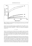
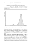
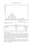
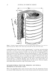
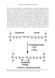
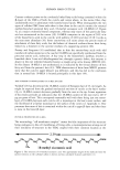
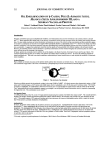
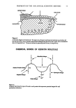
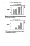
HUMAN HAIR CUTICLE 27 A-layer. Thus, together with an external layer of covalently linked fatty acid (see below), this seems likely to comprise the thin membranous sacs (so-called epicuticle) specifically raised from the cuticular surfaces of all mammalian keratin fibers when they are treated with dilute chlorine water (19,20). Isodipeptide-rich proteins also comprise thin, in- soluble intracellular envelopes in other keratinized structures such as the stratum cor- neum (21). Rice et al. (22) have reported TEM observations of mammalian hairs, from which most proteins had been extracted with hot mixtures of sodium dodecyl sulphate and dithiothreitol, and found isopeptide-rich proteins as a thin intracellular envelope for all the hair's cells, including the cortex. Thus all the cells of the hair shaft seem to possess these thin insoluble envelopes, and it is highly likely on this basis that the whole intracellular surface of each cuticle cell, and not merely the A-layer, possesses such an envelope. The suggestion has been made (23) that, on the assumption the majority of the cystine residues of the A-layer proteins are engaged in intermolecular crosslinking, this will render the structure tough and fitting in its role of protecting the hair surface from mechanical insult. Such a highly crosslinked protein, one anticipates, would swell little in water (as compared with other structures in the hair shaft). It can be expected to possess a relatively high indentation hardness and a very high modulus of elasticity as compared with the components of the hair cortex. One also expects the A-layer to fracture under applied tensile stress at strain levels significantly less than for the hair as a whole, and indeed this could be the focus for catastrophic circumferential fractures through the cuticle sometimes seen in individually stretched hairs or in hair tresses multiply combed (24). There is the additional possibility that the A-layer and accom- panying exocuticle undergo fracture at relatively low levels of strain in the bending of the cell sheet. This could be an advantage in ensuring that only small pieces are shed from the cuticle scale edges by frictional interaction, thereby helping to extend the cuticle's lifetime in protecting the main bulk of the hair shaft. The A-layer provides support for the rigid attachment of covalently linked fatty acids at the hair's surface (see below), which undoubtedly aids the frictional performance at this surface. THE EXOCUTICLE This sheet-like component, which again is proteinaceous, smoothly abuts the A-layer, but its other side is highly sculptured. Its thickness, accordingly, varies between ca. 100 and 300 nm. Under the TEM the exocuticle appears to be amorphous. Electron histo- chemical experiments have shown it to be particularly rich in cystine, i.e., at a higher concentration than in the proteins of the hair cortex but not as high as in the A-layer (5). Differential protease digestion of physically isolated human hair cuticle has been used to derive an amino acid analysis for the proteins of the exocuticle (6). Approximately 20% of the amino acid residues were as •/5-cystines. That the structure is relatively devoid of isodipeptide crosslinks is indicated by the relatively high speed of its digestion with papain/dithiothreitol, a reagent that otherwise only slowly dissolves the adjacent isodi- peptide-rich A-layer. Individual proteins have not yet been specifically isolated from the exocuticle, and so we don't know how many different types there are, or their amino acid sequences or conformation. The proteins can be expected to belong to the classes of high- and ultrahigh-sulphur proteins commonly identified in polyacrylamide gel electropho- retic separations from solubilized whole fibers (10,12).

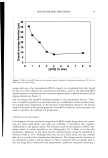
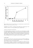
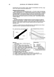
28 JOURNAL OF COSMETIC SCIENCE There is little doubt that the high level of crosslinking afforded by the exocuticle's cystine content will provide considerable strength and, in combination with the over- lying A-layer, will be fitting in the role of protecting the other surface of the hair from mechanical damage. THE ENDOCUTICLE The distinctive feature of this sheet-like proteinaceous subcomponent of highly variable thickness within each cuticle cell (ca. 50 to 300 nm) is the low concentration of cystine it contains as compared with the other major constituents of the cuticle. Reliable amino acid analyses have been obtained for the whole endocuticle and some of its substructures following differential digestion of physically isolated cuticle from human hair with a selection of proteases (6). Elevated levels of acidic and basic amino acid residues are in accord with moderately intense staining of the endocuticle seen under the TEM in sections stained with uranyl acetate/lead citrate (UA/LC) and phosphotungstic acid (PTA), respectively (5). The endocuticle seems to consist of the cellular debris remaining and pushed to one side as the A-layer and exocuticle have formed. Most TEM staining regimes show it to be of moderately coarse but irregular structure, even showing occa- sional pockets of cystine-rich material, which are evidently minor incursions from the adjacent exocuticle. Light microscope observations by Kassenbeck (25), in which he separated cuticle by high-temperature treatments with mixtures of ethylene glycol and toluene sulphonic acid, demonstrated the presence of a disk-like unit in the center of each cell sheet, undoubtedly defining the location of the cell's effete nucleus within the endocuticle. Using systematic protease digestions, Swift and Bews (6) were able to show that the endocuticle consists of at least three chemically and morphologically distinct protein components of relatively low cystine content, but all containing higher levels of acid and basic amino acids than other cuticle structures. These were identified as the chromatin of the effete nucleus, the non-chromatin components of the nucleus, and the non-nuclear debris. That these structures are so readily digested by non-reductive pro- tease treatments points to isodipeptides and disulphides being absent from them. Intact proteins have not been isolated, and so nothing is known of their amino acid sequences and likely conformations. The high basic and amino acid content of the endocuticle, coupled with the relative absence of intermolecular crosslinks in the form of cystine or isodipeptides, are likely to render this component mechanically soft and susceptible to significant swelling by water. These are in sharp contrast to the expected behavior of the other major compo- nents of the cuticle, the A-layer and exocuticle (see above). Atomic force microscope (AFM) observations of human hair (26) and wool (27) have indicated a considerable increase in the surface-scale step heights of the fibers as they are taken from a dry to a water-wet state. Most of this change is probably accommodated by swelling of the endocuticle and little by the cuticle's other structures. It is conjectural whether the endocuticle confers any special advantages to the owner's hair. One possibility is that it might offer some protection by providing a cushion beneath the tougher outer layers of each cuticle cell from forces impacting the hair surface. THE INNER LAYER This relatively minor lameliar subcomponent sits between the endocuticle and the CMC
Purchased for the exclusive use of nofirst nolast (unknown)
From: SCC Media Library & Resource Center (library.scconline.org)