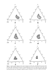
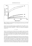
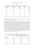
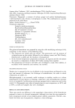
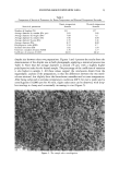
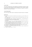
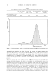
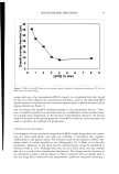
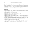
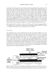
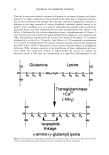
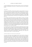
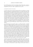
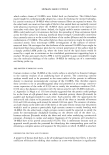
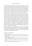
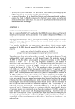
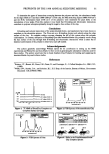
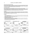
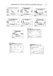
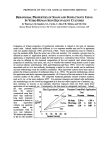
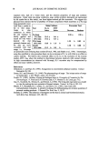
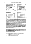
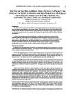
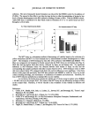
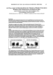
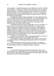
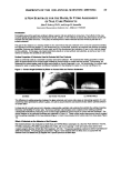
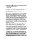
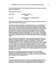
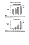
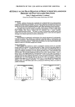
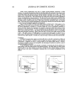
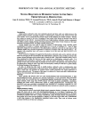
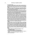
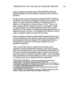
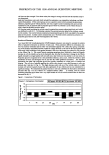
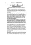
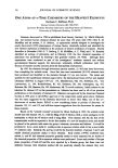
70 JOURNAL OF COSMETIC SCIENCE Figure 2: Results of Adhesion Testing on Three Nail Enamels Nail Enamel Adhesion on Glass Substrate (a) Product I (on Glass) (b) Product 2 (on Glass) (c) Product 3 (on Glass) Nail Enamel Adhesion on VITRO-NAILS Substrate (j) 3X View of Enamel J Summary , r I • r r Ii F I: D B A L J 1 In Vitro Evaluation of Nail Enamel Durability (Resistance to Chipping) Resistance to chipping was evaluated on nail enamel films (8 mils) drawn on VITRO-NAILS substrate. The films were allowed to dry for 2 hours, cut into 2 cm x 5 cm strips using a paper cutter and mounted between two rigid plastic holders (Figure 3). Twelve different enamels were tested simultaneously. The plastic holder was subsequently clamped to secure the strips and then mounted on the rod of a Tekmar RW 20 mixer. The rotating rod was then positioned horizontally above the hard textured target surface, such that the bottom edge of the strips would strike the target once per rotation. The rod was then rotated at 100 rpm for 6 hours. Upon completion, the VITRO-NAIL strips were visually inspected for chipping and flaking of the enamels. Figure 3: Nail Enamel Chipping Results: VITRO-NAILS 6-Hour Durability Test ( The nail enamels tested, varied significantly in the degree of chipping and flaking observed. The best performing products showed minimal damage after 6 hours (Figures 3e, 3d), while the worst performing products exhibited pronounced chipping and flaking at the 6 hour time point (see magnified views of Figures 3j, 3f). Validation versus actual "in use" testing is required to confirm that these results translate into consumer perceivable differences in wear and chipping. (f) 3x View of Enamel F We have developed a synthetic nail substrate that exhibits the wetting properties, thickness and flexibility of human nails. Preliminary in vitro adhesion and in vitro wear results look promising. These methods need to be refined and validated versus actual "in use" testing to confirm their usefulness as tools to accelerate the development of superior formulations. References 1. Paul N. Gardner Company Inc., Pompano Beach, FL. (d) Product I (on VITRO-NAILS) (e) Product 2 (on VITRO-NAILS) (f) Product 3 (on VITRO-NAILS) Our results indicate that all three nail enamels adhere well to glass (Figures 2a, 2b, 2c). Interestingly, when these same nail enamels were tested on a substrate with wetting properties similar to human nails, it was possible to observe performance differences among the three products (Figure 2d, 2e, 2f). These preliminary adhesion results using the new substrate are encouraging validation versus actual "n use" testing is required to confirm that these results translate into consumer perceivable differences.

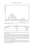
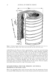
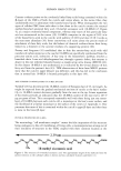
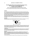
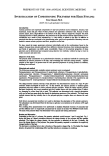
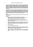
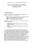
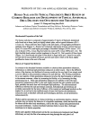
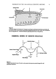
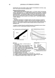
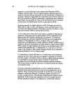
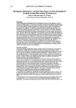
PREPRINTS OF THE 1998 ANNUAL SCIENTIFIC MEETING 71 HUMAN NAIL AND ITS TOPICAL TREATMENT: BRIEF REVIEW OF CURRENT RESEARCH AND DEVELOPMENT OF TOPICAL ANTIFUNGAL DRUG DELIVERY FOR ONYCHOMYCOSIS TREATMENT Jonas C.T. Wang and Ting Sun, Ph.D. Johnson and Johnson Topical Formulation and Drug Delivery Technology Resource Center, Johnson and Johnson Consumer Products, Skillman, New Jersey, USA Biochemical Properties of the Nail The human nail plate is composed of approximately 25 layers of flattened, keratinized cells fused into a dense, hard yet slightly elastic plate, with a typical thickness of 0.5 - 1.0 mm (1). These cells have their origin in the nail matrix, a living highly proliferative epithelia tissue (Figure 1). Human nail compared with human stratum corneum has less lipid (1% versus 20%) and higher percentage of disulfide linkages (10.6% versus 1.2%) also is thicker (750 p versus 30p) but holds less water (25% versus 300%) (ref. 2-4). The high disulfide bond content confers toughness to the nail (3). Nail consists of three layers (Figure 1): (i) hard dorsal nail plate 0.Smm thick in finger nails and 1.3mm for toe nails, (ii) nail bed of noncornified soft tissue, and (iii) nail matrix which is the thick, highly proliferative tissue at the root of the nail. History of Topical Nail Delivery In contrast to the abundant literature available on chemical skin penetration enhancers, information on chemical nail penetration enhancers is rather scarce. Due to the different nature of the barriers, the likelihood of success would not be very high if one simply used a proven-effective skin penetration enhancer for nail delivery. The working mechanism for a vast majority of skin penetration enhancers involves the lipid domains or pathways in the stratum corneum., either by increasing the fluidity, or by increasing the drag partitioning into it. These skin penetration enhancers are unlikely to have the same penetration enhancement effect on the nail simply because the nail contains much less lipid, probably has much less well-developed lipid pathways. The aforementioned differences between the nail and stratum corneum, both physical and chemical, are probably responsible for the lack of efficacy of the topical nail antifungal products on the market (5), as well as for the ineffectiveness of some well known skin penetration enhancers, such as dimethyl sulfoxide and homologous alcohols, on nail permeation enhancement (6). Walters and his associates (2, 6, 7) discovered that, as a permeation barrier, a human nailplate does not mimic the behavior of a lipophilic membrane, which has been the case for almost all the other body membranes, such as the skin, vaginal and gastrointestinal mucousal membranes. Instead a hydrated nail plate behaves more like a hydrogel membrane in its barrier properties. Mertin and Lippold (7,8,9) pointed oat that for onychomycosis treatment (nail fungal infections), not only the flux of an antimycotic drag through the nail plate is of
Purchased for the exclusive use of nofirst nolast (unknown)
From: SCC Media Library & Resource Center (library.scconline.org)