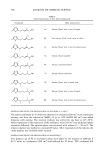
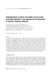
484 JOURNAL OF COSMETIC SCIENCE the vehicle but also drastically on the composition/internal structure of the phase, which may hamper drug diffusion in the vehicles (1-3). Recently, the use of peptide and protein domains with amphipatic sequences for drug and gene delivery is getting increasing attention (4). The basic domain of human immunodeficiency virus type I (HIV-1) transactivator of transcription (TAT) protein was reported to possess the ability to traverse biological membranes efficiently in a process termed "protein transduction" (5-7). Furthermore, TAT peptide, chemically attached to various proteins, including horseradish peroxidase, [3-galactosidase, and ovalbumin, was able to deliver these proteins to various cells and even in mice tissues, with high levels in the heart, lung and spleen (8,9). Although the actual mechanism of TAT has not yet clearly been established, common structural features of TAT include the presence of basic amino acids (arginine and lysine) as well as the ability to adopt an alpha helical conformation (7). Parathyroid hormone (PTH) is a peptide hormone, a long chain containing 84 amino acids. Although its principal activity is calciotropic, published investigations have re- vealed that it also shows lipolytic activity in human adipose tissue (10,11). Recently, PTH-derived tripepride (GKH, glycine-lysine-histidine) was reported to stimulate [3-adrenergic receptor coupled to adenylyl cyclase via the stimulatory Gs protein, an increment of cAMP production that leads to activation of protein kinase A and phos- phorylation of hormone-sensitive lipase (HSL), resulting in glycerol release by fat cells (12). The short fragment of GKH that is nonpermeable into the cell was successfully attached to TAT (9-polylysine) in order to have both lipolytic and penetration effects on the skin, without affecting the lipolysis activity of GKH. In this study, we investigate the possibility of TAT-GKH as delivery vehicles without the loss of the lipolytic effect of GKH through TAT-GKH fusion, and discuss its usefulness as a cosmetic ingredient in slimming products. MATERIALS AND METHODS PREPARATION OF TAT-GKH PEPTIDE TAT-GKH peptide (KKKKKKKKK-GKH) was prepared with an automated peptide synthesizer (Applie Biosystem 433A) by using standard solid-phase fluoenylmethoxy- carbonyl (Fmoc) chemistry with HATU as the peptide-coupling agent. Cleavage from the resin was achieved by using a mixture of trifluoroacetic acid (TFA/H20/ ethanedithiol/phenol/thioanisole). Removal of the solvent gave a precipitant that was triturated with cold diethyl ether. The crude mixture obtained was centrifuged, then removed by decantation, and the resulting orange solid was purified by HPLC (Shimazu LC-8A) in the linear gradient of 28-30% of CH3CN in 0.1% TFA for 15 min. The product was isolated by lyophilization and characterized by MALDI-TOF mass spec- trophotometry (Perseptive Biosystems Voyager linear mass spectrophotometer) by using ot-cyano-4-hydroxy-cinnamic acid as a matrix. The purity of the peptide was 95% as determined by analytical grade HPLC (13). CELL CULTURE 3T3-L1 cells (mouse preadipocytes) were obtained from American Type Culture Col- lection (Rockville, MD), and were grown to confluence in a basal medium, Dulbecco's

SKIN PENETRATION ENHANCEMENT BY TAT-GKH 485 Modified Eagle's medium (DMEM), supplemented with 10% fetal bovine serum (FBS) and penicillin/streptomysin (all from Life Technologies, Gibco, BRL). The medium was changed every two to three days. After confluence, the 3T3-L1 cells were differentiated into adipocytes by the addition of 2 lag/ml of insulin, 2 laM of dexamethasone, and 111 lag/ml of methyl-isobutylxanthine (all from Sigma) to the basal medium. After 48 h, the medium was replaced by 10% FBS basal medium, DMEM, containing only 2 lag/ml insulin, for ten days, during which time the cells developed into mature adipocytes with typical histologic appearance. LIPOLYSIS MEASUREMENTS The lipolytic effect, i.e., the degradation of triglycerides to glycerol and fatty acids, was evaluated by determining the quantity of glycerol released by the cells. Lipolysis was measured as the percent of glycerol in TAT-GKH-treated adipocytes vs glycerol in nontreated adipocytes (basal lipolysis) using Krebs-Ringer bicarbonate buffer containing 1% FBS (pH = 7.4) as incubation medium. After the adipocytes were washed with phosphate buffer saline (PBS, pH = 7.4), the TAT-GKH (final concentration 10 -4 M - 10 -7 M) dissolved in the incubation medium was added to the adipocytes and incubated at 37øC for 2 h. The lipolytic agent isoproterenol (Sigma) was used as a positive control at 10 -6 M. After incubation, the reaction mixture was centrifuged at 100 g at room temperature for 30 sec to separate the medium and the adipocytes. The glycerol content of the incubation medium was determined using a colorimetric assay (GPO-Trinder, Sigma) (14). ANIMALS Male Sprague-Dawley rats weighing 370-420 g and 8-9-week-old female hairless mice weighing 27-33 g (DaeHan Biolink, Taejeon, Korea) were housed in a temperature- controlled room (22 ø + 2øC) and subjected to a 12-h light/dark cycle. Animals had free access to laboratory food and water and were carefully handled. IN VITRO LIPOLYSIS ON ADIPOCYTES FROM RATS For in vitro experiments, rats were sacrificed by cervical dislocation after an overnight fast, and epididymal adipose tissue was immediately removed. Isolated fat cells were obtained by collagenase digestion (1 mg/ml, 37øC) in Krebs-Ringer bicarbonate buffer containing 3.5 g/100 ml of bovine serum albumin (BSA) and 0.6 mM of glucose at pH 7.4 (KRBA), under continuous vigorous shaking (90 cycles/min) according to the method of Rodbell (15). Fat cells were filtered through nylon mesh and washed three times with the same incubation buffer (KRBA), to eliminate the stroma-vascular frac- tion and collagenase A. Measurements of lipolytic activity were performed by incubating isolated adipocytes in 200 pl of KRBA buffer with continuous gentle shaking (30 cycles/min). After two hours of incubation with TAT-GKH (10-4--10 -7 M) at 37øC, the reaction was stopped with ice and an aliquot (50 pl) was taken to determine glycerol release in the incubation buffer by the calorimetric assay (14). Basal lipolysis was determined in the absence of TAT-GKH. The metabolic activity was expressed as micromoles of glycerol released per milligram of total lipids, which were determined gravimetrically after solvent extraction (16).
Purchased for the exclusive use of nofirst nolast (unknown)
From: SCC Media Library & Resource Center (library.scconline.org)