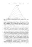
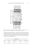
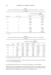
486 JOURNAL OF COSMETIC SCIENCE CYTOTOXICITY ASSAY Preconfiuent 3T3-L1 preadipocytes were seeded in 96-well plates at a density of 5 x 10³ cells/ml/well. The same doses of TAT-GKH were added to the culture medium. The cytotoxicity was evaluated by the MTT [3,(4,5-dimethylthiazol-2-yl)-2,5- diphenyltetrazolium bromide] test (17) and by measuring formazan formation spectro- photometrically at 570 nm. SKIN PERMEATION STUDIES Vertically assembled Franz-type diffusion cells (Microette transdermal diffusion system, Hanson Research Corporation, Chatsworth, CA) were used for in vitro skin permeation experiments. The system consisted of Franz-type diffusion cells with an effective diffu- sion area of 1.776 cm 2 and a receptor volume of 7.0 ml, an autosampler, and a cell drive system with rpm controller. The fundamental experiments were performed according to the method given in our previous report (18). Briefly, the excised skin of female hairless mouse was obtained from 8-9-week-old, 27-33-g animals. The dermal side of the skin was soaked in buffer with 15% ethanol solution containing 5 mM of phenylmethylsul- fonyl fluoride (PMSF) for 12 h at 10øC to inhibit the enzyme. The skin was mounted on a diffusion cell, and the receiver compartment was filled with 7 ml of 50 mM PBS with 15% ethanol containing 5 mM PMSF and maintained at 32øC by circulating water within a jacket around the lower chamber. PMSF was used to inhibit the enzyme and 15% ethanol was used to dissolve the PMSF. The ethanol concentration had no effect on the penetration of peptides, as reported by Ghanem et al. (19), who showed that ethanol at low levels (25%) had little or no effect on pore pathways. A 20% ethanol solution containing 1% TAT-GKH (w/v) and 1% GKH (w/v) was applied in the donor com- partment and was uniformly distributed with a micropipette on the skin surface (100 pl). The receptor fluid was mixed by a magnetic stirrer throughout the experiment and was collected from the receiver compartment at predetermined time (every 12 h after sample application) and replaced by fresh fluid. At the end of the experiment (24 h after sample application), the receptor fluid was collected and the donor compartment was washed with 500 pl of ethanol three times. After completion of the preset time (24 h), the skin samples were taken out of the diffusion cells. The skin was homogenized by 4 ml of PBS to extract TAT-GKH and GKH. After filtration on Millex filter FG (pore size: 0.2 pm, Millipore), the solutions were assessed by HPLC. Five hundred microliters of the receptor fluid withdrawn from the receiver compartment at predetermined times was treated with 10 pl of TFA. Following centrifugation (13,000 rpm), the amounts of TAT-GKH and GKH in the supernatants were determined by analytical HPLC. HIGH-PERFORMANCE LIQUID CHROMATOGRAPHY (HPLC) The HPLC consisted of a solvent delivery pump (600 pump, Waters Co., MA), a C•8 column (HP ODS hypersil 5 !•m, 4.6 x 150 mm, Hewlett Packard, Germany), a 486 UV detector (Waters), and a data process system (Waters Millennium). The TAT-GKH was analyzed with the mobile phase of distilled water in 0.1% TFA. TAT-GKH was ana- lyzed with the mobile phase of 15% acetonitrile in 0.1% TFA and at a flow rate of 1 ml/min. Absorbance at 210 nm was measured for the assay of these peptides. The

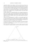
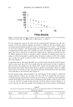
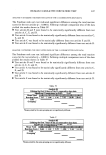
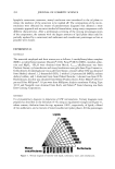
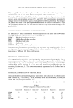
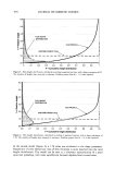
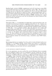
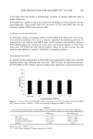
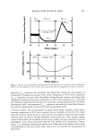
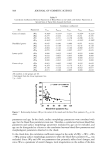
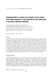
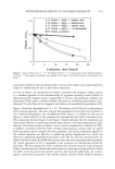
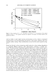
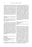
SKIN PENETRATION ENHANCEMENT BY TAT-GKH 487 retention time was 4.5 min for.GKH. The retention time was 12.3 min for TAT-GKH. The temperature of the column was kept at 40øC. STATISTICAL ANALYSIS Values are given as means + SEM of (n) separate experiments. Differences in value were statistically analyzed using a two-tailed two-sample t-test. RESULT AND DISCUSSION IN VITRO LIPOLYSIS IN CULTURED ADIPOCYTES Due to the fusion of TAT into GKH, it was necessary to confirm whether TAT-GKH, like GKH, still had the same lipolytic effects or not. To compare the lipolysis effects of TAT-GKH with those ofGKH, 3T3-L1 differentiated adipocytes were incubated in the presence and absence of various doses of TAT-GKH and GKH at 37øC for 2 h. Lipolysis in 3T3-L1 differentiated adipocytes was estimated by determining the amount of glyc- erol released into the medium as a result of lipolysis on triglyceride. The lipolytic agent isoproterenol was used as the positive control to confirm the absence of technical errors in this experiment and induced high lipolytic effects (data not shown). The effects of increasing concentrations of TAT-GKH and of GKH on glycerol production in adipo- cytes are depicted in Figure 1. TAT-GKH induced approximately 37.6% maximal lipolysis at 10 -5 M, compared with basal lipolysis. Both TAT-GKH and GKH repre- sented similar lipolytic effects at the same concentration and also induced lipolytic effects in a dose-dependent fashion. IN VITRO LIPOLYSIS IN ADIPOCYTES ISOLATED FROM RATS 3T3-L1 differentiated adipocytes contain considerable triglyceride in much smaller lipid storage droplets than mature primary adipocytes, which contain large unilocular lipid 6O ,-- 5O ß 30 • 20 o '10 10-4 10-• 10-• 10-7 [] GKH ß TAT-GKH Concentration (mol/L) Figure 1. Lipolytic effects of TAT-GKH in 3T3-L1 differentiated adipocytes, compared with those of GKH. Values are mean _+ SEM (n = 6), expressed as percent (%) vs basal lipolysis for glycerol and are significantly different from those for basal lipolysis.
Purchased for the exclusive use of nofirst nolast (unknown)
From: SCC Media Library & Resource Center (library.scconline.org)