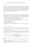
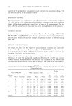
38 JOURNAL OF COSMETIC SCIENCE TOPICAL FORMULATIONS All formulations were prepared on a weight/weight basis. There were three formulations used in this study, a simple solution of ot-TAc (5%) in isopropyl myristate (IPM), an emulsion formulation of ot-TAc (5%), and a solution of ot-T (1%) in IPM. The com- position of the emulsion formulation is given in Table I. RECEPTOR FLUID In the metabolism study the receptor media must serve two important functions, viz., maintaining the viability of skin tissue and ensuring the solubility of the prodrug and its metabolite. Dulbecco's modified phosphate buffered saline (DMPBS) was used to maintain the tissue viability (21,22). As compounds that are essentially insoluble in water (such as the ot-T derivatives) may not partition freely from excised skin into an aqueous receptor fluid, bovine serum albumin (3%) was added to DMPBS (21,23). Fresh deionized water was used to prepare the buffer, and the solutions were adjusted to pH 7.4 with 10% w/v NaOH prior to use. ANIMAL TISSUE AND PREPARATION Fresh viable micro-Yucatan pig skin was obtained from Charles River Laboratories (Wilmington, MA). The pig skin was cut into squares of 10 x 10-cm 2 pieces and placed in Tupperware © containers filled with DMPBS. The skin was then set on cool packs and used for the metabolism experiments within one day of its arrival. Upon receipt the fresh skin was gently washed with a 1% (w/w) mild soap and deionized water. A 250-300- pro-thick layer of the skin was cut from the surface with a Padgett Electrodermatome TM instrument (Padgett Instrument Co., Kansas City, MO). The dermatomed skin was used the same day and was cut into 10-mm circular pieces with a brass punch and placed epidermis-side-up in Bronaugh diffusion cells. DOSING Finite dosing was used to simulate actual use conditions in all the ], vitro permeation and metabolism experiments. The smallest volume of the formulation required to obtain complete and uniform coverage of the diffusion cell surface area (0.636 cm 2) was de- Table I Composition of •-TAc (5 %) Emulsion Formulation Concentration (% w/w) Ingredient ot-TAc •-TAc 5 Diisopropyl adipate 7.5 Mineral oil 7.5 DEA-cetyl phosphate 2 Carbomer 0.3 Diazolidinyl urea 0.3 Water q.s. 100

PERMEATION AND METABOLISM OF o•-T and o•-TAc 39 termined to be 5 pl, corresponding to a weight of about 4 mg. After application the preparation was uniformly spread on the stratum corneum (SC) side of the skin with the help of a glass rod, and the tip of the rod was washed into a vial containing 2 ml acetonitrile in order to account for the material lost on spreading. With this technique the exact amount of material applied on the skin surface was determined. IN VITRO SKIN PERMEATION/METABOLISM METHODOLOGY A flow-through system was used for conducting in vitro permeation experiments. The total system consisted of a receptor fluid reservoir, a variable flow rate peristaltic pump, Cassette © (Manostat, New York, NY), a circulating water bath, Lauda © (Brickman Instrument Co., Westbury, NY), two cell-holding heating blocks, 14 Teflon © flow- through diffusion cells, and a Retriever IV fraction collector (ISCO Inc., Lincoln, NE) to collect effluent fractions over the adjusted time period. Each diffusion cell had an inner diameter of 9 mm and a surface area of 0.636 cm 2 exposed to the receptor fluid. The receptor fluid was pumped at a flow rate of 1.5 ml/h from the reservoir to the diffusion cells placed in the holding blocks. The skin surface temperature was maintained at 32øC by adjusting the circulating water bath temperature to 39øC (24). Effluent from diffu- sion cells was collected directly into glass scintillation vials. The experiments (four replicates for each formulation) were stopped at time intervals of 2, 6, 12, and 24 hours. SKIN TREATMENT At the conclusion of the experiment at each time point in the kinetic study, the donor compartment was washed thrice with 1 ml of acetonitrile. Washes were collected and analyzed by HPLC for the amount of drug remaining on the surface. Washed skin samples were removed from the cells. The tape-stripping technique was used to separate the stratum corneum (SC) from the rest of the epidermis to get an estimate of material remaining in the barrier layer of the skin. In this technique, seventeen strips of the drug-treated side of the skin, using a 3M Scotch TM tape, were taken as two + 15 strips. The first two strips represented the drug superficially adhering to the surface (and so included in the wash), and the next 15 strips represented the drug recovered from the SC. To each of the two strips and 15 strips about 15 ml of n-hexane was added exactly. Both these strips were shaken in a wrist-action shaker for 45 minutes at the end of which the mixture was filtered and injected into the HPLC. The remainder of the skin was placed in plastic culture tubes, 5 ml of ice cold DMPBS was added, and the skin was homogenized (Polytron homogenizer, Switzerland) for five minutes until a buff-colored suspension was obtained. Absence of chunks of skin was ensured. This suspension was extracted three times each with 5 ml of n-hexane. Each extraction involved shaking the mixture on a wrist action shaker for 45 minutes. After extraction a 45-minute centrifugation process helped to separate the n-hexane and DMPBS layers, and the upper n-hexane layer was carefully removed with a pipette and pooled together into 30-ml glass tubes. This procedure was repeated three times, each time shaking for 45 minutes and centrifuging for 45 minutes for each skin sample. The pooled hexane layer was evaporated under vacuum. Acetonitrile (2 ml) was added to this mixture, vortexed to ensure complete mixing, and the solution was filtered and injected into the HPLC column. The whole extraction procedure had been validated previously
Purchased for the exclusive use of nofirst nolast (unknown)
From: SCC Media Library & Resource Center (library.scconline.org)