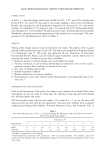
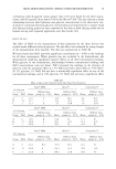
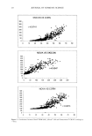
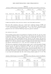
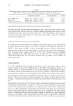
2000 ANNUAL SCIENTIFIC MEETING 81 the instruments based on the p-values to be significant. It may be the result of the small base size (10) of the one-hour test. Table 4: Correlation between the one-hour change in reading and the two-week improvement in skin grade. Device Regression equation of Score R-squared P-value Reduction (SR) on 1 hour reading NOVA DPM SR = 0.0025X + 0.69 0.917 0.042 Skicon 200 SR = 0.0024X + 0.81 0.969 0.015 Corneometer CM 825 SR = 0.0209X + 0.76 0.8427 0.082 There was also good correlation between the instrumental readings taken 16 hours post treatment and the actual skin grades with all three instruments (data not shown) CONCLUSIONS Salt does effect the electrical readings after application of moistunzers to the skin but the there is still a good correlation between glycerin concentration and the electrical readings with all three instruments tested Results obtained with 10 panelists, 1 hour after treatment were predictive of moisturizing efficacy in the two week, twenty panelist test with all three instruments.. Our results show that single application tests can be predictive of longer-term results with humectant based molstunzers and that electrical measurements correlate well with skin grades. REFERENCES 1 E. Berardesca, EEMCO guidance for the assessment of stratum corneum hydrat•on electncal methods, Skin Res Technol, 3, 126-132 (1997). 2 H Tagaml, "Measurement of electrical conductance and impedance," in Handbook of non-invasive methods and the skin, J Serup and G Jemec, Eds. (CRC Press Inc, Boca Raton, 1995), pp 159-164 3 M Loden, B•ophys•cal methods of providing objective documentation of the effects of mo•stur•z•ng creams, Skin Res Technol, 1, 101-108 (1995). 4 A O Barel, P. Claws and B. Gabard, "In v•vo evaluabon of the hydratlon state of the skin measurements and methods for claim support," m Cosmet/cs Controlled Efficacy Studies and Regulation, P Eisner, H F Merk, and H I Ma•bach, Eds. (Spnnger-Verlag, Berlin, 1999), pp 57-80 5 J W. Fluhr, M Gloor, S. Lazzenm, P Kleesz, R Grieshaber and E Berardesca, Comparative study of five instruments measuring stratum corneum hydration (Corneometer CM 820 and CM 825, Skicon 200, Nova DPM 9003, DermLab) Part I In wtro, Skin Res. and Technol., 5, 161-170 (1999) 6 P Clarys, A. O Barel and B Gabard, Nonqnvas•ve electncal measurements for the evaluation of the hydrabon state of the skin comparison between three conventional instruments- the Corneometer, the Sbcon and the Nova DPM, Skin Res Technol, 5, 14-20 (1999) 7 W Courage, "Hardware and measuring principle corneometer," In Bloengineenng of the Skin' Water and the Stratum Corneum, P Eisner, E Berardesca, and H I Ma•bach, Eds (CRC Press Inc, Boca Raton, 1994), pp 171-175 8. B Gabard and P Treffel, "Hardware and measunng pnnc•ple the NOVA DPM 9003," •n Bioengineering of the Skin Water and the Stratum Corneum, E B P Eisner, H.I. Maibach, Editor (CRC Press, Inc, Boca Raton, 1994), pp 177-195. 9 H TagamL "Hardware and measuring pnnc•ple sbn conductance," •n B/oengineenng of the Skin Water and the Stratum Corneum, P Eisner, E Berardesca, and H I Ma•bach, Eds. (CRC Press Inc, Boca Raton, 1994), pp 197-203 10. D L Bissett and J. F McBnde, Sbn conditioning w•th glyceroh J. Soc Cosmet Chem, 35(11), 345-350 (1984).

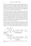
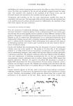
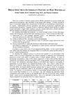
82 JOURNAL OF COSMETIC SCIENCE COSMETIC APPLICATIONS OF A WOUND HEALING PEPTIDE Karl Lintner, Ph.D., Philippe Mondon, Ph.D., Oliver Peschard, Ph.D. and Claire Mas-Chamberlain, Ph.D. Serderma, Le Perray en Yvelines, France Introduction Wound healing is a very complex process, the mechamcs of which have not yet been completely unravelled. Although some general features are common to the wound repair mechanisms in different organs and tissues, we shall focus here on the activities observed in skin tissue, which in any case, when we think of a "wound", comes to mind first. Wound healing process What happens when we cut our skin? The first step in damage control is to stop the potentially deadly loss of blood: coagulation occurs in the micro- vessels and fibrin is deposited to obdurate any holes or openings. This occurs through a cascade of protein activation steps. Peptides cleaved from circulating proteins during this process are not just by-products, but have their own purpose and activity. They are often chemotacfic, that is, they attract cells (platelets, leucoc3•tes, macrophages and fihroblasts) to the site of the wound. Some peptides then act on the damaged cells and provoke their death (apoptosis, a safety feature), others act on the remaining cells and on those chemically attracted cells to stimulate them into the synthesis of new tissue. Indeed, once the blood flow has been taken care of, inflammation (more or less pronounced depending on the size of the wound) sets in: Fibroblasts first secrete proteolytic enzymes to help clean out the wound, swelling due to increased cell density and blood flow occurs. After the damaged tissue has been removed and oedema has been circumscribed, the process of reconstruction of the conjuncfive tissue begins by deposition of collagen fibres and glycosaminoglycanes. The covering of the wound site by epithelial cells also initiates the synthesis of matrix macro-molecules in the fibroblasts • What makes the fibroblasts change their activity from "lytic" to "synthetic"? It appears that an extemal signal, like a local hormone, a messenger molecule that interacts with the fibroblast cell membrane receptors, induces this change. And what kind of signal might that be? Obvious candidates are small signal peptides, those that are released from the degradation process of the macromolecules. This feedback mechanism is a logical consequence of the wound-healing cascade and confirms that mother Nature is economical: use every waste product possible for some beneficial purpose! Some of these peptides, fragments of the degraded macromolecules, have now been isolated, identified and synthesised in larger quantities for further investigation. If we consider that the ageing process (induced by sun exposure and the ensuing inflammatory reactions, the free radicals, the upregulation of elastolysis and collagenolysis) is a slowly occurring wound (the "injuries" of time, Horace, Odes II1), then the use of these signalling "wound healing peptides" in cosmetics seems worth pursuing. We investigated the biological and cosmetic activity of a model peptide Ala-g-His (camosine) and three peptides that are fragments of elastin (Val-Gly-Val-Ala-Pro-Gly =VGVAPG, chemotactic), collagen I (central sequence: Gly-His-Lys =GHK) and procollagen I terminal sequence (Lys-Thr-Thr-Lys-Ser =KTTKS). For reasons of time and space we shall discuss only the latter one here. Material and Methods Peptides were synthesized by classical Merrifield solid phase methods. In vitro and ex vivo measurements of collagen and GAG synthesis are described elsewhere (2,3). Results and Discussion Skin diffusion In order to use biologically active peptides in cutaneous application at concentration levels that are safe, effective and nevertheless economical, optimum skin diffusion must be assured. For this purposes it is necessary to modify the peptide sequence in order to improve cutaneous substantivity and diffusion. We therefore added a palmitoyl chain to the N-terminal amino acid and then tested the in vitro, ex vivo and in vivo activity of the Pal- KTTKS molecule. In a previous model study we had compared the skin diffusion (using radiolabels) of a dipeptide Ala-His with its palmitoylated equivalent Pal-Ala-His.
Purchased for the exclusive use of nofirst nolast (unknown)
From: SCC Media Library & Resource Center (library.scconline.org)