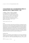
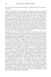
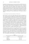
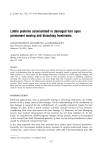
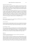
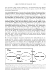
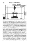
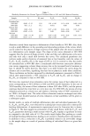
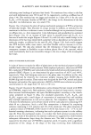
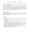
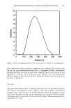
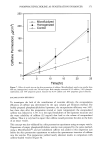
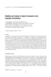
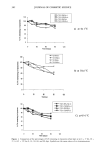
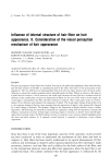
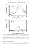
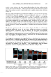
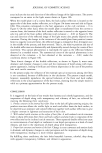
PHOSPHATIDYLCHOLINE AS PENETRATION ENHANCER 365 20 - 18 16 14 12 0 500 600 100 200 300 400 Size(nm) Figure 1. Vesicle size distribution of sample 1 (2.0% phosphatidylcholine, 0.5% caffeine, 10% propylene glycol). DETERMINATION OF VESICLE SIZE Samples were diluted with distilled water and the vesicle size distributions were deter- mined by photon correlation spectroscopy (Zetasizer 3000HS, Malvern Instruments Ltd, Malvern, UK). The measurements were conducted in a CONTIN mode, and the inten- sity-based mean vesicle sizes were reported. ENCAPSULATION EFFICIENCY The encapsulation efficiency of the lipid vesicles was determined by the spin column gel filtration method (14). One gram ofSephadex G50 was swollen in 10% propylene glycol aqueous solution for three hours and washed two times with the same solution. A small piece of glass wool was inserted in the bottom of a 10-ml syringe and the prepared gel was poured into the syringe. To separate the encapsulated caffeine from free caffeine in the solution, 1 ml of the vesicle dispersion was added and the column was centrifuged for two minutes at 2000 rpm and again with 1 ml of 10% propylene glycol solution. The concentration of eluted caffeine was analyzed by HPLC. Free caffeine molecules were

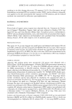
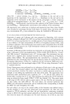
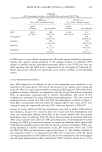
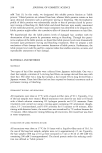
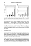
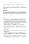
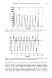
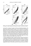
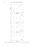
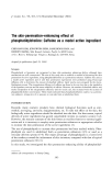
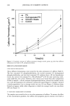
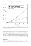
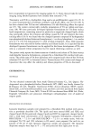
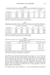
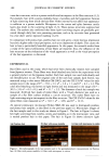
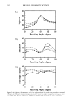
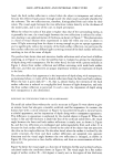
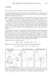
366 JOURNAL OF COSMETIC SCIENCE 20 18 16 14 12 to 10 • 8 o 600 100 200 300 400 500 Size(nm) Figure 2. Vesicle size distribution of sample 2 (2.0% hydrogenated phosphatidylcholine, 0.5% caffeine, 10% propylene glycol). bound to the Sephadex gel and remained in the column. The ratio of the amount of entrapped caffeine to the total amount of caffeine in the dispersion was calculated. IN VITRO SKIN PERMEATION EXPERIMENT In vitro skin permeation tests were performed with abdominal skin of a female hairless guinea pig (strain IAF/HA-hrBR) using Franz diffusion cells (Lab Fine Instruments, Korea). The receptor compartments of the Franz cells were filled with 5 ml of PBS solution (pH 7.4) and constantly stirred by a magnetic bar at 600 rpm. The excised skin samples were mounted with the stratum corneum sides facing the donor compartments, with a contact area of 0.636 cm 2. Each 300 i•1 of the lipid vesicle dispersion to be tested was applied to donor compartments, and the temperature of the system was maintained at 32øC by a circulating water jacket. The entire content of each receptor solution was withdrawn at 6, 12, and 24 hours after the lipid dispersion was applied, and the receptor compartments were refilled with fresh PBS solution to maintain the sink condition. The receptor solution samples were assayed for caffeine by HPLC.
Purchased for the exclusive use of nofirst nolast (unknown)
From: SCC Media Library & Resource Center (library.scconline.org)