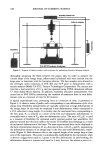
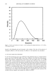
EFFECTS OF CAPPARIS SPINOSA L. EXTRACT 327 [LOOH]p,.i. = AT9 o - AT5/eTsEc = ^5/TsE % inhibition = ([LOOH]R.• ' -[LOOH]•nH[LOOH]}t.•. ) X 100 where R.I. -- radical initiator AT9 o and AT5 = absorbance at the end and at the beginning of the experiment A'•9 o and A'T5 = absorbance at the end and at the beginning of the experiment in the presence of the antioxidant • (molar extinction coefficient of conjugated dienes) = 26, 100 ß 400 M -• cm-• Ts• c (time, sec)= 5,100 [LOOH]R.i.: hydroperoxide concentration after addition of the radical initiator alone and [LOOH]• n = hydroperoxide concentration after addition of the antioxidant. All experiments were carried out in triplicate and repeated at least three times. Results were calculated as percentage decrease with respect to control values, and mean inhibi- tory concentrations (IC5o) were evaluated by using the Litchfield & Wilcoxon test. IN VIVO EVALUATION OF THE PHOTOPROTECTIVE EFFECTS OF LECS Preparation of aqueous gels. Carhomer gels were prepared by dispersing, with constant stirring, carhomer (Carbopol 934 0.8% w/w as final concentration) and triethanolamine (0.9% w/w as final concentration) in a suitable amount of water:ethanol (60:40) solution so as to obtain a 2% final concentration of LECS or of tocopheryl acetate (TOC), used as reference substance. The resulting gels were stored at room temperature for 24 h under air-tight conditions prior to use. A gel formulation without active compounds was used as control (CONTROL). Instrument. UVB-induced skin erythema was monitored, as previously reported (6,21), by using a reflectance visible spectrophotometer, X-Rite model 968 (X-Rite Inc., Grand- ville, MI), having 0 ø illumination and a 45 ø viewing angle. The instrument was cali- brated with a supplied white standard traceable to the National Bureau of Standard's perfect white diffuser. The spectrophotometer was controlled by a computer, which performed all color calculations from the spectral data by means of a menu-driven suite of programs (Spectrostart, X-Rite Inc.) supplied with the instrument. Reflectance spec- tra were obtained over the wavelength range of 400-700 nm using illuminant C and a 2 ø standard observer. Protocol. Experiments were performed on six volunteers of both sexes in the age range of 25-35 years. The volunteer subjects were fully informed about the nature of the study and the procedures involved. No subject was known to exhibit abnormal sensitivity to sunlight, or was taking any medication at the time of the study. The experiments were performed under standardized room conditions (22 ø + 2øC and 40-50% relative hu- midity) after a resting time of 15 min. Skin erythema was induced by UV-B irradiation using an ultraviolet lamp, model UVM-57 (UVP, San Gabriel, CA). This source emits in the range of 290-320 nm with an output peak at 302 nm. The flux rate measured at the skin surface was 0.80 mW/cm 2. For each subject, the minimal erythemal dose (MED) was preliminarily determined, and an irradiation dose corresponding to double the MED was used throughout the study. For each subject, six sites on the ventral surface of the forearms were defined using a circular template (1 cm 2) and were demarcated with permanent ink. Skin sites were exposed to UV-B irradiation, and then the gel formulations containing tocopheryl acetate or LECS (100 mg) were spread uniformly on the four sites (each gel formulation two times) by means of a solid glass rod. For each volunteer, two of the six sites were

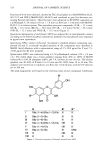
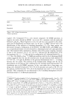
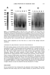
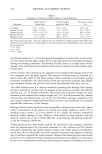
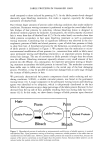
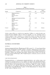
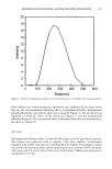
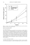
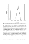
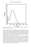
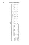
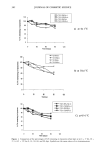
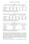
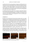
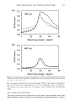
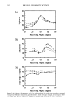
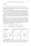
328 JOURNAL OF COSMETIC SCIENCE used as controls (applying the gel formulation without altive compoundskinEach site, after gel application, was occluded for 3 h, using Hill Top chambers (Hill Top Research, Inc., Cincinnati, OH), to prevent any loss of material from the skin surface. After the occlusion period, the chambers were removed and the skin surfaces were washed to remove the gel and allowed to dry for 15 min, after which the induced erythema was monitored for 58 hours by reflectance spectrophotometry. From the reflectance spectral data obtained, the erythema index (E.I.) was calculated using an equation similar to that reported by Dawson et al. (28): E.I.=100 log--+l.5 log +log -2 log --+log T2o TZgo where I/R is the inverse reflectance at a specific wavelength (560, 540, 580, 510, and 610 nm). E.I. baseline values were taken at each designed site for gel formulation before UV-B irradiation, and they were subtracted from the E.I. values obtained after UV-B exposure at each time point, to obtain AE.I. values. For each site, plotting AE.I. vs the time the area was under the curve (AUOo_sg) was computed using the trapezoidal rule. To better compare the efficacy of the different gel formulations tested, the percentage inhibition of UV-B skin erythema (PIE) was calculated from AUOo_sg values using the following equations: A UC(c ) - A UCcc ) Inhibition (%)= x 100 AUC(c) where AUC(c ) is the area under the response-time curve of the sites treated only with the gel formulation without active compounds (CONTROL), and A UC(•) is the area under the response-time curve of the sites treated with LECS or TOC gel formulations. Sta- tistical analysis was performed by using Student's t-test. RESULTS AND DISCUSSION TOTAL PHENOL CONTENT AND CHEMICAL COMPOSITION OF LECS The total phenol content expressed as rutin equivalents, determined by the Folin- Ciocalteau method, was 65.13 _+ 5.53 mg/g (see Table I). TLC analysis of LECS revealed the presence of flavonols and hydroxycinnamic acids as major constituents. A Sephadex LH-20 column, followed by purification using reversed- phase HPLC gave pure compounds I-VIII (Figure 1), giving a more complete profile of the flavonol content of C. spinosa buds. Flavonols were identified by electron spray mass spectra (ESIMS) and extensive NMR analyses as (see Figure 1) kaempferol-3-O-o•-L- rhamnopyransyl-( 1 - 6)-[o•-L-rhamnopyranosyl-( 1 - 2)- •-D-glucopyranoside] (I) (29), kaempferol-3-O-o•-L-rhamnopyranosyl-(1- 6)-•-D-glucopyranoside (III (17), querce- tin- 3-O-o•-L-rhamnopyranosyl-( 1 - 6)- •-D-glucopyranoside (V) ( 18,30-32), and quer- cetin (VII) and kaempferol (VIII) (33). These represent the most common flavonoids occurring in C. spinosa, and rutin has been reported to be the most abundant in the floral button and leaves (18,32). Compounds II, kaempferol-3-O-o•-L-rhamnopyranosyl-(1 - 2)-•- D-glucopyranoside, and IV, quercetin-3-O-o•-L-rhamnopyranosyl-(1- 6)-[o•-L-rhamnopy- ranosyl-(1-2)-•-D-glucopyranoside], are here reported for the first time in the genus
Purchased for the exclusive use of nofirst nolast (unknown)
From: SCC Media Library & Resource Center (library.scconline.org)