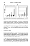
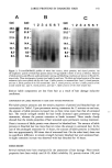
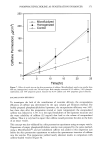
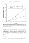
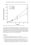
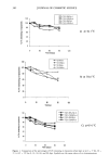
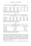
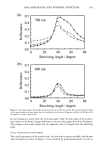
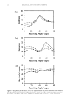
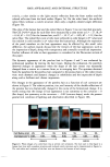
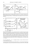
EFFECTS OF CAPPARIS SPINOSA L. EXTRACT 325 graphs were plotted showing a linear relationship between concentration versus peak areas for both reference compounds. LECS was redissolved in MeOH and analyzed under the same chromatographic condi- tions used for compounds III and V. The total concentration of the quercetin derivatives (compound IV, V, and VII) was expressed as quercetin-3-0-rutinoside (rutin) equiva- lents the total concentration of the kaempferol derivatives (compounds I, II, III, VI, and VIII) was expressed as kaempferol-3-0-rutinoside equivalents. Quantitative HPLC analysis of hydroxycinnamic acids. An aliquot (50 mg) of LECS was dissolved in dimethyl formamide 2 ml of 2 N NaOH was added, and the solution was stored in the dark for 4 h. Then the mixture was acidified with 2 N HC1 to pH 2 and extracted three times with 10 ml of ethyl acetate. The three ethyl acetate fractions were evaporated under vacuum, and the residue was recovered with 1.5 ml of methanol. Chromatography was performed on a Hypersil ODS column (particle size: 5 l•m 25 cm x 4.0 mm i.d. Perkin-Elmer, Norwalk, Connecticut). The mobile phase was water:tet- rahydrofuran:acetic acid, 70:18:2. The flow-rate was set at 1.0 ml/min. Each sample was filtered prior to injection using a Millex HV13 filter (Waters-Millipore Corporation, Milford, MA), and an aliquot (20 1•1) was injected into the HPLC apparatus. Detection was effected at 300 nm. The retention times were 8.9 min for caffeic acid, 10.56 min for ferulic acid, 13.48 min for p-cumaric acid, and 28.55 min for cinnamic acid. The peak assignments were based on the retention times of a single compound. Cali- bration graphs were plotted showing a linear relationship between concentrations versus peak areas for all compounds. IN VITRO ANTIOXIDANT AND RADICAL SCAVENGING ACTIVITIES OF LECS Bleaching of the free radical 2,2,-diphenyl-l-picrylhydrazyl (DPPH test). The antiradical activity of LECS was determined using the stable 2,2-diphenyl-1-picrylhydrazyl radical (DPPH ø) and the procedure described in the literature (23,24). In its radical form, DPPH ø has an absorption band at 515 nm that disappears upon reduction by an antiradical compound. An aliquot (37.5 lal) of a water:dimethylsulphoxide (90:10) so- lution containing different amounts of LECS was added to 1.5 ml of daily prepared DPPH ø solution (0.025 g/1 in methanol) the maximum concentration of the extract employed was 200 lag/ml. As reference compounds, ot-tocopherol and ascorbate (two well-known antioxidants) and quercetin and kaempferol (the flavonoids contained in LECS) were employed these reference antioxidants were employed also because they possess different reduction potentials (25,26). An equal volume (37.5 l•l) of the vehicle alone was added to control tubes. Absorbance at 515 nm was measured on a Shimadzu UV-1601 UV-visible spectrophotometer 20 min after starting the reaction. The DPPH ø concentration in the reaction medium was calculated from a calibration curve analyzed by linear regression. The percentage of remaining DPPH ø (%DPPH'REM) was calcu- lated as follows: %DPPH'wE M = [DPPH']T/[DPPHø]o x 100 where T is the experimental duration time (20 min). The percentage of remaining DPPH ø against the antioxidant concentration was then plotted to obtain the amount of antioxidant necessary to decrease the initial concentra-

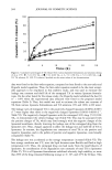
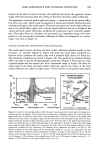
326 JOURNAL OF COSMETIC SCIENCE tion by 50% (mean scavenging concentration, SC5o). All experiments were carried out in triplicate. Protective effect against UV radiation-induced peroxidation in liposomal membranes (UV-IP). The protective effect of LECS against UV-induced peroxidation was evaluated on phosphatidylcholine (PC) multilamellar vesicles (20). Briefly, 1.0 ml of liposome suspension (in a glass flask with a 3-cm 2 exposure surface area) was exposed to UV- radiation from a 15-watt Philips germicidal lamp (254 nm) for 1.5 h. Exposure was given at 10 cm from the lamp, at room temperature. An aliquot (50 l•l) of a water:di- methylsulphoxide (90:10) solution containing different amounts of LECS was added to the system the maximum concentration employed was 200 t•g/ml. Quercetin and kaempferol, the flavonoids contained in LECS, were tested as reference compounds. An equal volume (50 l•l) of the vehicle alone was added to control tubes. The malondial- dehyde (MDA) concentration in the mixture was measured by using a colorimetric assay kit (Calbiochem-Novabiochem Corp., La Jolla, CA). All experiments were carried out in triplicate and repeated at least three times. Results were expressed by a decrease in percentage with respect to control values, while mean inhibitory concentrations (IC5o) were calculated by using the Litchfield & Wilcoxon test. Oxidation of linoleic acid in LA/DPPC LUVs (LP-LUV test). The method consists in the spectrophotometric determination of the accumulation of products (conjugated dienes) of peroxidation, induced by the water-soluble peroxyl radical generator 2,2'-azobis(2- amidinopropane)hydrochloride (AAPH), of linoleic acid (LA) in mixed dipalmitoylphos- phatidylcholine/linoleic acid (DPPC/LA) unilamellar vesicles (LUVs) (23,24,27). Mul- tilamellar liposomes (MLVs) were obtained by freshly prepared chloroform-methanol (1:1, v:v) concentrated solutions of DPPC and LA (molar ratio 1:0.125). The solvents were removed under nitrogen in a rotoevaporator, and the resulting film was kept overnight under vacuum to remove the residual solvents. Liposomes were prepared by adding 0.9% NaCI aqueous solution to the film, then heating at a temperature above that of the gel-liquid crystalline phase transition (60øC) and vortexing three times for 1 min, after which the samples were shaken for 1 h in a water bath at 60øC to homogenize the liposomes. LUVs were prepared by submitting the previously prepared MLV dispersion to extrusion through 100-nm polycarbonate membranes (Avestin Inc.) in an extruder system (LiposoFast Basic ©, Avestin Inc.). An aliquot (6 l•l) of a water:dimethylsulphoxide (90:10) solution containing different amounts of LECS was added to 1.2 ml of LUV suspension (21 mg DPPC/ml) as reference compounds o•-to- copherol (a lipophilic antioxidant) and ascorbate (a hydrophilic antioxidant) and quer- cetin and kaempferol (the flavonoids contained in LECS) were tested. Then the mixture was incubated for 20 min at 37øC in a shaking water bath, after which the peroxyl radical generator AAPH was added to the suspension to obtain a final concentration of 10 t•M. The oxidation was carried out at 37øC (below the transition temperature of DPPC/LA LUVs) under air. At given times (5-90 min), 120 l•l aliquots of the reaction mixtures were withdrawn and added to 1 ml of methanol. The accumulation of LOOH formed from LA was determined spectrophotometrically by measuring the absorbance of the samples at 233 nm. The ratio of oxidation-induced change in absorbance, with and without addition of antioxidant, was used to calculate a percentage inhibition of oxi- dation (100% corresponding to complete protection and 0% corresponding to no dif- ference from control) by the following equations:
Purchased for the exclusive use of nofirst nolast (unknown)
From: SCC Media Library & Resource Center (library.scconline.org)