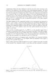
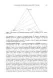
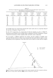
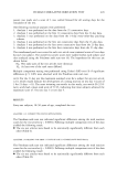
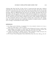
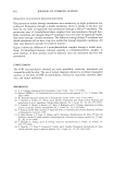
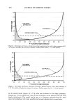
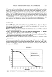
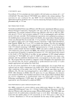
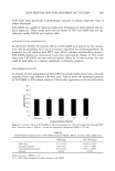
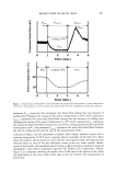
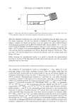
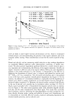
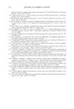
500 JOURNAL OF COSMETIC SCIENCE (4-7). There have also been many reports about the effects of cosmetics on the mor- phology of the cutaneous surface using these methods (6,8,9). In previous studies, we analyzed replicas of the skin obtained from eight sites (forehead, glabella, corner of the eye, upper eyelid, lower eyelid, nasolabial groove, corner of the mouth, and cheek) on the faces of 136 healthy Japanese females (aged 18-83 years) using a three-dimensional surface morphology measurement system, and we evaluated age-associated changes (10). The results indicated that age-associated increases in the depth of wrinkles were the greatest in the corner of the eye, followed by the glabella, the corner of the mouth, the nasolabial groove, the upper eyelid, the forehead, the lower eyelid, and the cheek. We hypothesized that wrinkles are formed under the influence not only of ultraviolet rays but also of other factors. However, how this surface morphology is related to other parameters of skin properties has scarcely been evaluated except for its relationship with the physiologic state of the cutaneous surface (11,12). Concerning cutaneous blood flow, there have been a few reports on its age-associated changes in human facial skin (13-15) and other studies concerning its relationships with primary stimuli such as transepidermal water loss (TEWL) and erythema (16,17). Al- though decreases in cutaneous blood flow are generally supposed to have adverse effects on the skin, little is known about how such changes affect cutaneous properties in normal skin. Thus, clarification of the effects of cutaneous blood flow on the three- dimensional morphology of the skin surface, particularly wrinkles formed on the face, is important. In this study, therefore, we evaluated the relationship between the surface morphology and blood flow of facial skin by measuring the cutaneous blood flow in 40 elderly subjects, quantifying the three-dimensional morphology of the skin surface using rep- licas, and examining correlations between blood flow and morphological parameters. MATERIAL AND METHODS SUBJECTS AND DESIGN OF THE STUDY The subjects used in this study were 40 healthy Japanese women aged 60-77 years, who lived in the suburbs of Tokyo and gave informed consent. The subjects washed their faces with liquid face-washing agents (Kao Corporation, Tokyo) prior to each measurement. MEASUREMENTS USING INSTRUMENTS Measurement of blood flow and analytical parameters. Blood flow was measured by the method of Nagashima et al. (15,18,19). Each measurement was performed using an integration-type skin blood laser-Doppler flowmeter with a temperature loading instru- ment (ILD-T, Kao Corporation, Tokyo, Japan) at two points, on the forehead and on the cheek. The measurement on the cheek was made on the zygomatic bone to ease attaching the probe. After the subject had been acclimated at 25øC and 50% RH for 15 minutes, the blood flow was measured with the subject in the spine position. The blood flow was monitored serially for one minute at a probe temperature of 30øC, for two minutes with cooling at a probe temperature of 10øC, and for two minutes after the end of cooling. Figure 1 shows the definitions used for blood flow parameters. Parameter Frest represents the mean resting skin blood flow for 30 seconds at a sensor temperature of 30øC (mV),

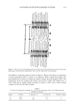
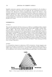
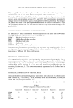
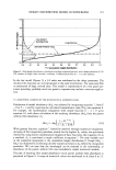
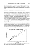
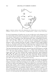
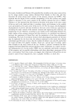
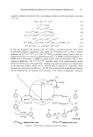
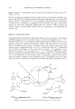
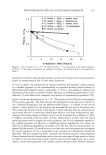
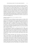
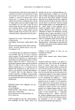
BLOOD FLOW IN FACIAL SKIN 501 Frest Finter-L Fpost-L cooling 10øG o o o 0 I 2 3 4 5 Time Imin.} 30øc cooling 10øC free m 30 o • 10 0 1 2 3 4 5 Time (min.} Figure 1. Typical chart of blood flow. The blood flow was measured for 30 seconds at a sensor temperature of 30øC, for two minutes at 10øC, and for two minutes after the low temperature setting was canceled. parameter Fmi n represents the minimum skin blood flow during the two minutes of cooling after changing the setting of the sensor temperature to 10øC (mV), parameter Finter_ L represents the mean skin blood flow during the two minutes of cooling after changing the setting of the sensor temperature to 10øC (mV), parameter Fma X represents the maximum skin blood flow between the end of the cooling period and the end of the measurement (mV), and parameter Fpost_ L represents the mean skin blood flow between the end of cooling period and the end of the measurement (mV). Collection of replicas and 3D measurement of replicas. Each subject entered a room with a constant temperature of 20øC and a constant relative humidity of 40-50% (10). After about 60 minutes, she lay down on a bed with her eyes lightly closed, and replicas were obtained from six areas of the face (forehead, corner of the eye, lower eyelids, cheeks, corner of the mouth, and nasolabial groove) using a rubber precision impression material (hydrophilic vinyl silicon impression material: GC Exafine, GC Corporation, Tokyo). Figure 2 is a schematic showing the replica sites. Collection of the replica on the cheek was performed on the lower cheek rather than at the site used for the measurement of blood flow.
Purchased for the exclusive use of nofirst nolast (unknown)
From: SCC Media Library & Resource Center (library.scconline.org)