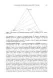
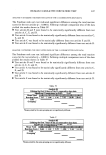
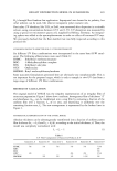
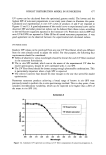
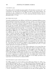
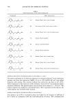
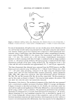
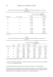
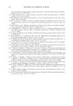
PHOTODEGRADATION BY RUTILE-BASED PIGMENTS 515 compounds undergoing degradation via either pathway or both (15). In this technical note the use of phenol and salicylic acid as model molecules is extended to a wider range of commercial inorganic pigments, with the aim of evaluating their overall photocata- lytic activity. EXPERIMENTAL We tested four coated rutile-based pigments, with Aldrich uncoated rutile as a control. The pigments used in this work are listed in Table I, together with their particle diameter (maximum of the distribution function of particle diameters, approximated with a Gaussian, as measured in reference 14). It was not possible to measure the particle size of the pigments coated with stearic acid (A and C) because they poorly dispersed in water. The poor water dispersion was also observed during photodegradation experi- ments, and thus the photocatalytic activity of these pigments is likely to be underes- timated in our experimental setup. Phenol (purity grade 99%) was purchased from Aldrich and salicylic acid (99%) from Carlo Erba. Aqueous suspensions of the pigments were obtained upon sonication with a Branson 2200 sonifier. The pigments (0.500 g 1-1) were irradiated in magnetically stirred Pyrex glass cells (diameter 4.0 cm, height 2.5 cm, suspension volume 5.0 ml) under a Solarbox (CO.FO.ME.GRA., Milan, Italy) equipped with a 1500-W xenon lamp and a 340-nm cutoff filter. Incident radiation in solution in the UVA region was 0.01 W cm -2 (14). The distance between the lamp and the Pyrex cells under the Solarbox (a closed irra- diation device with reflecting walls) was 21 cm. The Pyrex glass cells we used are shown in Figure 1. In the case of salicylic acid, the solution pH was about 3.5, and under such conditions the hydroxylation reactions are negligible when compared with the charge- transfer processes (17). Table I Pigments Used in Irradiation Experiments Partide Name Supplier Composition diameter (nm) (A) Micro titanium Tayca Corporation Rutile, coated with dioxide MT-100TV alumina and stearic acid (B) UV Titan M262 Variati & Co. Rutile, coated with alumina and dimethicone (C) Kemira UV Titan Variati & Co. Rutile, coated with M160 alumina and stearic acid (D) UV Sperse Biogenikko Rutile, coated with alumina and 1,3-butanediol (E) Titanium (IV) oxide Aldrich Rutile, uncoated 200 285 285 1400 (bimodal distribution) The particle diameters are taken from reference 14, where they were measured by means of a Coulter Model N4 MD laser-based submicron particle analyzer. The particle diameter could not be determined for the pigments coated with stearic acid since it was not possible to obtain a sufficiently homogeneous aqueous dispersion.

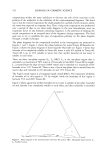
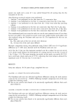
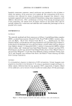
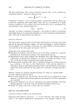
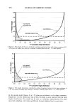
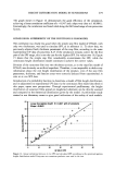
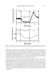
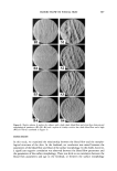
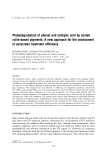
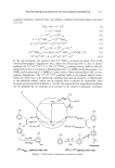
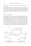
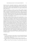
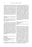
516 JOURNAL OF COSMETIC SCIENCE 25 ß Figure 1. Pyrex glass cell used for irradiation experiments (measures are given in mm). The level of the 5.0-ml suspension inside the cell and the magnetic stirring bar are also shown. After the scheduled irradiation time, each cell was withdrawn from the light source, and the whole suspension (5.0 ml) was filtered on Millipore HA syringe filters prior to analysis. Analysis of phenol and salicylic acid was carried out with a Merck-Hitachi chromatograph equipped with an RP-C18 LiChroCART column (Merck, 12.5 x 0.4 cm) packed with LiChrospher 100 RP-18 (particle diameter 5 pm). Elution was carried out with a 30/70 mixture of acetonitrile/phosphate buffer (total phosphate 0.050 M, pH 2.8), and the detection wavelength was 210 nm. Retention times at 1.00 ml min -1 flow rate were 3.65 min for phenol and 3.90 min for salicylic acid, the column dead time being 0.90 min. Before showing the experimental results it is useful to describe the photocatalytic processes at the basis of the degradation of both phenol and salicylic acid, the two model molecules used in this work. PHOTOCATALYTIC DEGRADATION PATHWAYS FOR PHENOL AND SALICYLIC ACID The irradiation of semiconductor oxides at wavelength }t hc/Ea,, where Ea, is the band-gap energy of the oxide, promotes electrons from the valence band into the conduction band, leaving holes in the valence band. In the case of titanium dioxide, the irradiation wavelengths giving the onset to these processes lie in the near-UV region (UVA or shorter). Photogenerated electrons (e-) and holes (h +) can either thermally recombine or migrate to the surface of the photocatalyst, where they can be trapped by surface and subsurface groups. Electrons are trapped as surface Ti TM species (often briefly named e-s•r f) and holes as surface TiIV-øOH (briefly øOHsurf or øOHad s) or subsurface Ti•v-o-ø-Ti•V (h+•ub_surf). The recombination between trapped electrons (Ti•n•urf) and trapped holes (Ti•v-øOH•urf and Ti•v-o-ø-Ti•V) is far slower than the direct recombi- nation between e- and h + in the semiconductor bulk (reaction 4), and reaction with molecules in solution is therefore possible. Ti TM species react with molecular oxygen to yield superoxide (they can also react with other electron acceptors in solution), while Ti•v-øOH•urf and Ti•v-o-ø-Ti TM can oxidize dissolved molecules, often up to complete mineralization. The Ti•v-o-ø-TiIV species are involved in electron-transfer reactions. On the contrary, the Ti•v-øOHsu•f groups usually act as hydroxylation agents with aromatic compounds, as hydrogen abstractors with aliphatic ones, and as electron acceptors with inorganic compounds (2). The cited processes are depicted in reactions 3-10, where S is
Purchased for the exclusive use of nofirst nolast (unknown)
From: SCC Media Library & Resource Center (library.scconline.org)