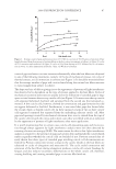
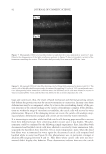
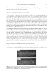
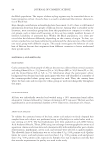
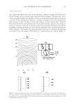
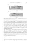
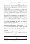
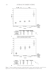
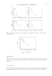
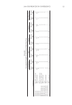
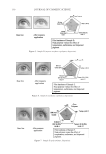
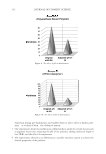
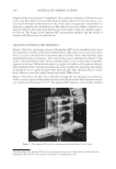
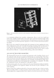
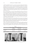
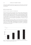
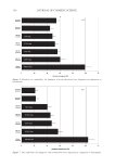
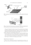
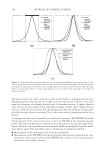
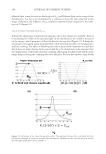
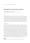
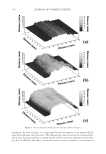
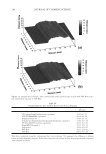
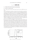
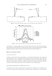
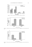
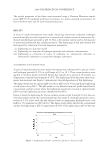
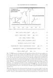
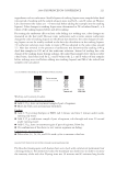
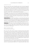
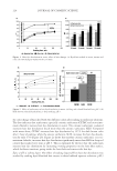
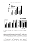
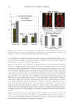
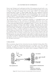
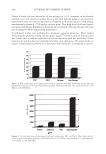
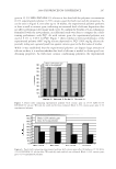
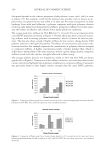
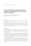
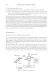
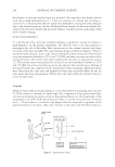
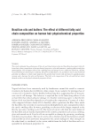
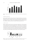
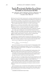
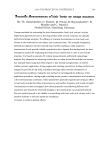
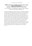
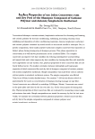
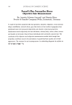
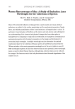
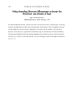
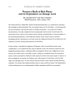
JOURNAL OF COSMETIC SCIENCE 86 EXPERIMENTAL PROTOCOLS The method used to study the formation of gaps and cavities within the cuticle sheath consisted in analyzing the light interference patterns (LIPs) produced by cuticle cells when seen under a powerful light by optical microscopy (4). As it has already been shown (4) cuticle cell decementation causes disruptions in the continuity of the cuticle sheath producing the inclusion of air in the formed cavities. Light interference patterns are then produced by a mismatch in the indexes of refraction between the cuticle cell layers and the air cavities as the light passes through. The presence of these defects within the cu- ticle sheath is thus revealed by the appearance of colorful patterns appearing at the hair surface. A similar phenomenon has been reported to occur with pores within the medulla of hair (5). MATERIALS AND INSTRUMENTS The light interference patterns were analyzed by microscopic analysis using a Hi-Scope KH-3000 equipped with a metal halide lamp. The hair used for testing thermal and mechanical damage was European virgin hair from International Hair Importers. For experiments requiring analysis of hair close to the root, virgin hair from 4 female donors was obtained from areas close the scalp. Hair from the same source was used for lipid extraction. Solvents used for oil and lipid extraction from hair were IPA and Hexane GC grade. Extraction of lipids from hair was made by immersing 1 g bundles of hair fi bers in 100 ml of solvent at room temperature conditions for periods of time ranging from min- utes to months. Analysis of the lipids extracted from hair was made by GC-MS after methylation. During solvent immersion single hair fi bers were selected and taken for microscopic anal- ysis at various time intervals. When needed, cycles of mechanical and thermal stresses were applied to single hair fi bers using a blow dryer or curling iron according to methods described elsewhere (6–7). The number of gaps and cavities appearing at the cuticle sheath of hair fi bers, either treated with solvents or damaged mechanically or thermally, were counted and plotted as a function of hair length. All defects producing patterns of light interference ranging in size from 0.1 to 5 microns were considered as gaps or micro- cavities. The oil used for deposition into these cavities was mainly Jojoba oil. The deposi- tion method consisted in immersing the hair fi bers in a solution of oil at 0.1% in IPA at RTC for three minutes. RESULTS GAPS AND CAVITIES FORMED BY MECHANICAL AND THERMAL STRESSES Microscopic analysis of hair fi bers taken from the various donors showed that the presence of gaps and cavities in people’s hair was pervasive. Figure 1 shows, for instance, that the average count of gaps and microcavities increases in regions of the fi ber distant from the root, while in areas close to the root the count is nil. Also, it was observed that the average

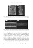
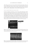
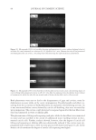
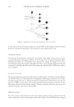
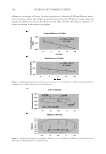
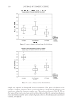
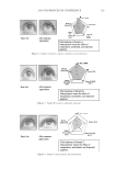
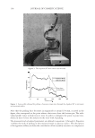
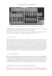
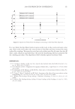
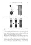
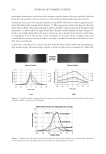
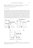
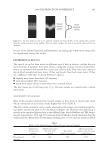
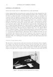
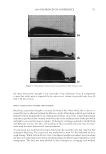
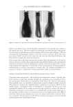
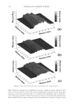
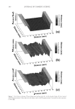
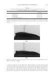
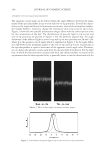
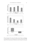
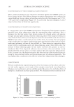
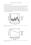
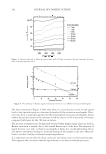
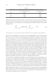
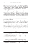
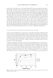
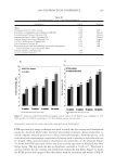
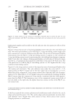
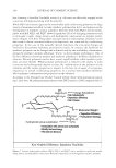
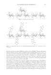
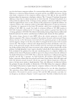
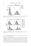
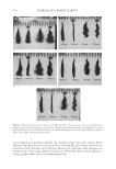
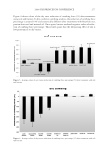
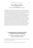
2008 TRI/PRINCETON CONFERENCE 87 count of gaps and micro-cavities increases substantially when the hair fi bers are subjected to any of the following treatments, namely: (i) Cycles of mechanical stresses, (ii) cycles of thermal stresses, or (iii) immersion in solvents (see Figure 1). It should be mentioned here that the average number of gaps and cavities found along the various hair fi bers was seen to vary strongly from subject to subject. The shape and size of defects giving rise to the appearance of patterns of light interference was observed to be dependent on the type of stresses applied to the hair fi bers. Cycles of mechanical extension and retraction usually led to the formation of extended gaps or large open cavities between the two top cuticle cells (see Figure 2). In most cases the top cuticle cells appeared deformed, buckled, and separated from the second one that remained ce- mented. A few cuticle cells, however, showed de-cementation and gap formation but did not appear deformed or buckled. Furthermore, it was noted that gaps also formed after the second, third, or fourth cuticle cell. In these instances stacks of two or three cuticle cells appeared cemented but separated from the underlying adjacent cuticle cell. Most gaps and openings created by mechanical extension were seen to extend from the tips of the cuticle cells towards the cortex and in most cases they were fi lled with air as indicated by the appearance of patterns of light interference after stress application. It is worth mentioning that in certain cases cuticle cells presenting gaps and microcavi- ties though always apparent by light interference analysis were not often detected by scanning electron microscopy (SEM). The main reason for this is that light interference analysis is sensitive to the presence of gaps and cavities deep underneath the cuticle sheath surface regardless whether the cuticle cells are buckled or not. Previous research has al- ready shown that these types of extended gaps involving cuticle cell decementation are formed by Poisson contraction stresses acting on the cuticle sheath as the hair fi bers are subjected to cycles of elongation and retraction (6). The cyclic radial contraction and extension of the hair fi bers during elongation produces cuticle cell cement breakage by fatigue, and also induces viscoelastic deformations on the whole cuticle cell, therefore, causing buckling and creating extended gaps or cavities fi lled with air. Figure 1. Average count of gaps and microcavities (15% SD) in a section of 50×50 μm as a function of hair length obtained from four groups of ten hair fi bers each after various treatments as follows: (a) After 20 cycles of 15% extension and retraction. (b) After 20 cycles of 10-s blow-drying at 65°C followed by 10 s of immer- sion in water. (c) After immersion in IPA for 3 min. (d) With no treatment.
Purchased for the exclusive use of nofirst nolast (unknown)
From: SCC Media Library & Resource Center (library.scconline.org)