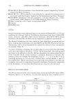
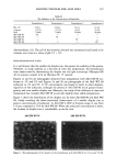
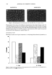
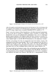
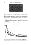
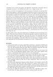
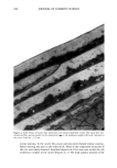
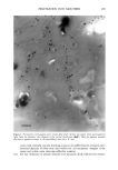
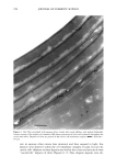
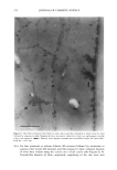
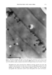
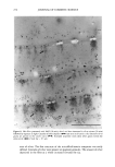
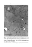
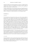
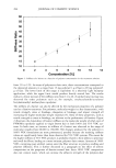
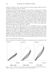
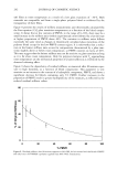
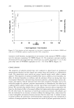
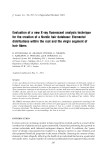
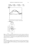
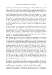
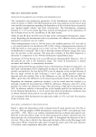
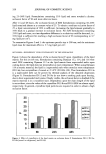
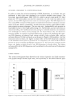
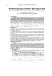
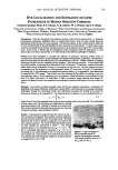
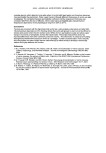
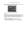
PENETRATION INTO HAIR FIBER 267 RESULTS 1. Medullated hair fibers viewed by light microscopy in the presence of water: With the dry fiber, the medulla was easily observed as a light or dark structure de- pending on the natural hair color. Observation was made easier with grey hair. Within one minute of the application of water, progressive changes in the optical appearance of the medulla could be seen. In localized areas the walls of the medulla changed in contrast relative to its previous dry state and relative to the cortex (Figure 1). 2. Using the typical combination of aqueous uranyl acetate (five minutes) and aque- ous lead citrate (three minutes) to treat whole fibers, all sections showed typical cuticle staining (Figure 2). This included intense staining of the endocuticle and the delta layer of the cmc. The exocuticle was more lightly stained. Virtually no stain was observed in the a-layer. All cortical cells showed obvious staining, particularly of the cell nuclear remnants and inter-macrofibrillar material. 3. Hairs immersed in aqueous silver nitrate, rinsed, dried, and then exposed to light: For hairs immersed for two minutes, little to no staining of the cuticle was observed. Contrast in the cortex was too low to observe a typical microfibril/matrix composite structure. Silver grains were evident on the melanin granules. For hairs immersed for ten minutes, a similar result was found, i.e., no definitive contrast in any of the hair structures. Extensive deposits of silver grains were present in the cortex cell membranes (Figure 3). All of the pigment granules showed fine deposits of silver. 4. For hair fibers immersed in aqueous silver nitrate and then made alkaline with NaOH, all hairs showed morphological staining typical of arnmoniacal silver ß . • . ., • . t30 • t60 •. . •. Figure 1. Single medullated hair immediately after addition of water (t o) and after 60 seconds (t6o). Light micrograph. Changes in the appearance of the medulla due to hydration can be seen along the individual walls of the medulla (•--,•).

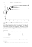
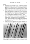
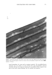
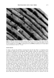
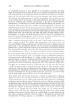
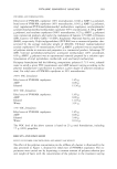
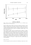
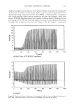
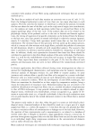
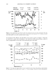
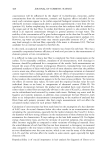
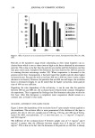
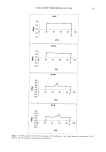
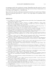
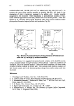
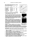
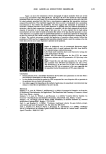
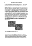
268 JOURNAL OF COSMETIC SCIENCE .. Figure 2. Cross section of normal hair pretreated with uranyl acetate/lead citrate. The stains have pen- etrated the fiber, giving contrast to the endocuticle (•--), cell membrane complex (•n) and structures in the cortex. Scale bar = 0.5 pm. nitrate staining. In the cuticle the a-layer and exocuticle showed intense staining. Sparse staining was seen in the endocuticle. None of the component structures of the cmc were easily observed. Localized deposits of silver were seen within the cell membrane complex of the cuticle (Figures 4, 5). The high-sulphur proteins of the
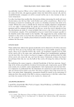
Purchased for the exclusive use of nofirst nolast (unknown)
From: SCC Media Library & Resource Center (library.scconline.org)