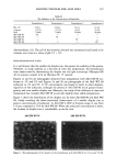
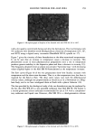
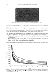
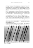
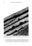
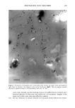
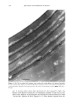
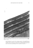
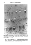
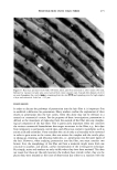
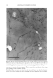
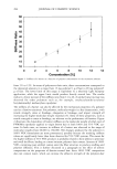
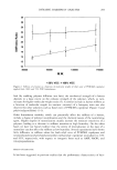
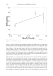
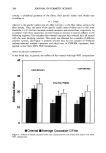
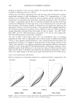
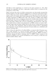
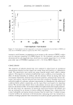
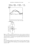
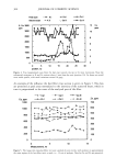
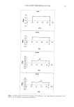
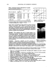
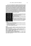
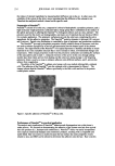
278 JOURNAL OF COSMETIC SCIENCE are cascade-like structures of silver granules. It is interesting to speculate that these might show penetration pores from the cmc into the sulphur-rich proteins of the cuticle cell. The deposits in the cmc show that chloride ions from the first treatment are retained within the cmc and interact with silver ions in the subsequent silver nitrate treatment. This indicates that both reagents have used the same pathway. Care must be exercised in the interpretation of the cascades, as the absence of deposits from adjacent areas may be due to nucleation and increased crystal growth in the areas of marked deposits. Alternatively, it is indicative of a localized reaction with the sulphur-rich proteins. It is clear from serial sections that these "cascades" are three-dimensional in structure and expand progressively from the crnc into the exocuticle. It is difficult to interpret deposits in the endocuticle, as these may indicate further penetration from the cmc or penetration along the endocuticle and retention in the known small sulphur-rich domains. The same findings were made close to the hair root where the cuticle cell edge would be intact. Interestingly, no cascades were found associated with the cuticle cell membrane bor- dering the cortex, although deposits of silver were seen in both the cortical cmc's and inter-macrofibrillar material abutting the cuticle cortex boundary. When considering observations made on the cortex of hairs treated with chloride and silver, caution is needed. The visually larger precipitates in the cortical cmc's and inter-macrofibrillar material could lead one to conclude that access to these structures is greater than to the macrofibrills. However, the cmc and inter-macrofibrillar material probably have a greater amount of free space vs the macro fibrils and, therefore, would be able to precipitate or develop more silver. The reduced contrast in the macro fibril does not exclude the presence of silver one would require further proof by elemental analysis. By combining the results of the above studies it becomes very evident that all structures in the hair can be accessed by small, water-soluble molecules, although the pathway to each structure may well be different. It is highly probable that access to the cortex is gained primarily via the cmc, whereas access to the cuticle cell is gained via the cuticle cell edge. The latter permits access into both the high-sulphur and low-sulphur com- ponents of the cuticle cell. However, imaging this event is very dependant on the choice of reagent and the affinity of ions for a particular site. It is not clear what role the "cascade" observations play in penetration into the cuticle cell. They are present at both root and tip, indicating they are independent of hair damage. In this way their presence and appearance are independent of the limited "pores" described by Swift (1) in weath- ered hair. Combined with the typical silver stains, here used e, bloc, it is now obvious that the high-sulphur proteins of the cuticle are easily penetrated. These studies show that the hair can present a number of compartments of differing affinity and capacity for penetrating molecules. It appears that the cuticle cell presents a large compartment, but one which would not deliver material, beyond its enveloping membranes, into the adjacent cortex. Viewing the cuticle as a definite, and open-ended, compartment helps to account for the rapid initial loss of hair dyes from the fiber with washing. Conversely, the cuticle cell membrane complex presents a small compartment allowing the transport of molecules across the cuticle to the cortex. One can only speculate on the capillary forces that might exist within the cmc lamellae between adjacent cuticle cells. The large compartment presented by the cortex, which readily expands in the presence of water, will act as a pulling force to increase flux along the cmc. Once an aqueous molecule enters the cortex, it will again be presented with compartments of varying capacity, i.e., macrofibril, nuclear remnant, and inter-

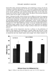
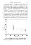
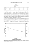
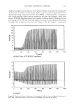
PENETRATION INTO HAIR FIBER 279 macrofibrillar material. What is very evident from these studies is that the medulla, at the center of the hair, the pigment granule within the body of the cortical cell, and the microfibril/matrix composite can all be accessed within one to two minutes or less at room temperature. It is also clear from these studies that the previous debate concerning the mode and route of penetration into the hair fiber needs further and careftA consideration. Much of the discussion is limited to considering the cuticle as a boundary structure that needs to be crossed. While this is true, it should also be viewed as a highly important compartment in its own right. Understanding the uptake and retention of materials into this com- partment and the transport of materials, via the cmc, across the cuticle depends on how the observations are made. It is most probable that access to the cortex is via the cuticle cell membrane complex. This multilamellar structure would favor transport parallel to the lamellae rather than normal to the lamellae. The suggestions by other authors that transport to the cortex occurs from the endocuticle would require molecules to traverse the cmc normal to its complex physicochemical lameliar structure. As yet we have no evidence for this route. CONCLUSION These studies have shown that aqueous molecules can be observed in all of the structures of the hair from one to two minutes after immersion in water-soluble reagents. Impor- tantly, these can be identified within the cuticle cell membrane complex and the bulk of the cuticle cell at the same time, indicating different but complementary pathways into the hair fiber. No evidence was found to suggest a diffusion pathway from the bulk of the cuticle cell, across the cmc, and into the cortex. Conversely, evidence was found for pathways from the cmc into the bulk of the cuticle cell along the whole of the hair fiber. By combining the correct reagents, a detailed knowledge of the hair structure, knowl- edge of the results and problems of product formulations, and, importantly, typical TEM stain results, one can build a more comprehensive view of penetration into the hair fiber. It is imperative to understand that the observation of a material in a particular structure does not always tell you how it got there. Nor does its absence from other structures say that it was never there. Importantly, penetration should be viewed as the movement of molecules from the outside of the fiber to various compartments in the fiber, rather than just from the outside of the hair to the inside of a cortical cell. ACKNOWLEDGMENTS I gratefully acknowledge Miss Patricia Goggin, Royal Holloway and Bedford College, for her technical assistance. REFERENCES (1) J. A. Swift, "The Detection of Pores and Holes in Hair by Electron Microscopy," in Hair Research for the Next Mi//eni•m, D.J.J. Van Neste and V. A. Randall, Eds. (Elsevier Science BV, Amsterdam, 1996), pp. 109-112.
Purchased for the exclusive use of nofirst nolast (unknown)
From: SCC Media Library & Resource Center (library.scconline.org)