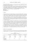
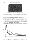
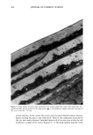
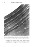
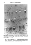
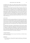
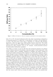
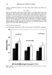
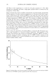
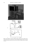
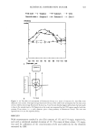
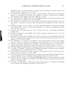
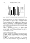
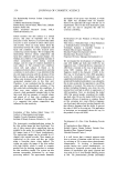
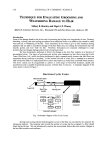
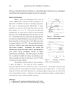
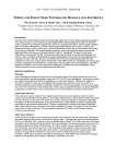
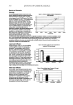
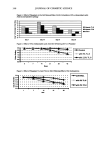
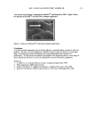
276 JOURNAL OF COSMETIC SCIENCE Figure 10. Root hair pretreated with NaCI (30 rain), dried, and then immersed in silver nitrate (30 rain) followed by exposure to light. Silver granules are present in the inter-macrofibrillar material (•) and occasionally within the macrofibrils. Subtle microfibril detail can be seen. Cascades are present in the inner cuticle cell (•,-•) but do not appear at the cuticle cortex boundary (,,•1•). Scale bar = 0.25 pro. structure or a reaction between reagents. This is a particular consideration when using static images to view a dynamic process. Medullated fibers in water are viewed in real time and hence can show the dynamic process of the hair swelling and water changing the optical properties of the medulla.

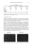
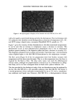
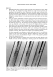
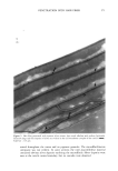
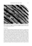
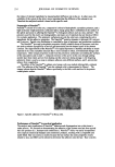
PENETRATION INTO HAIR FIBER 277 Indeed, it was possible to watch the wall structures of the medulla change progressively over a matter of seconds. This showed quite clearly that, with a highly active molecule such as water, one can achieve total access to the centre of the fiber in one minute or less. This suggests that for small, active molecules there is a rapid transport system operating in the fiber. However, it does not indicate a particular or preferential pathway. Using typical combinations of uranyl acetate and lead citrate on intact fibers, these studies showed that electron contrast, identical to that achieved with cross sections, could be achieved by pretreating the fibers. This indicates that the stains have complete access to each of the structures, e.g., the delta layer of the cmc, endocuticle, cortex cmc's, and macrofibrils. The relative absence of stain contrast in the exocuticle and a-layer would appear to indicate that these structures cannot be accessed. However, it is well documented that these structures do not show electron contrast when these stains are used on cross sections (7). One is, therefore, left with the question of "Did the stain get into the structure and leave during the rinsing step?" or "Is the structure inaccessible?" It is not possible to conclude from this approach whether the reagents entered via the delta layer and/or endocuticle it is only safe to say that the markers are retained in these structures even though these are the most likely penetration routes. Whole hairs treated with aqueous silver nitrate, rinsed, and exposed to light showed extensive silver deposition in the cortical cmc's and pigment granules. This indicates that silver has entered the cortical cell. Aqueous silver nitrate showed no conclusive pattern of contrast, i.e., there was no specific attachment to the high-sulphur proteins even though the previous result shows that molecules can easily enter many of the hair's structures. One is, therefore, left with the problem of "Is the silver present in the cuticle but invisible?" Hairs immersed in aqueous silver nitrate and then made alkaline to precipitate insoluble silver hydroxide showed strong staining of the high-sulphur proteins of the cuticle and cortex. In addition, they showed very distinct precipitates in the cell membrane complex of the cuticle. This shows that both aqueous silver and hydroxide ions were present in the cmc at the same time, indicating that the cmc is a pathway from the outside of the hair to the cortex. This also serves to show that deposits will only be observed when they have either an affinity for a structure or are made to stay where they are. In the case of silver nitrate alone, it is likely that it is removed from the cmc when the fiber is rinsed. Subsequent experiments where whole hairs were treated with only aqueous silver nitrate but without rinsing, drying, and exposure to light showed small amounts of silver precipitated in the cell membrane complex of the cuticle. In addition, some small deposits were evident in the a-layer. This further indicates that it is essential to find a method to localize markers of penetration, as absence of evidence is not evidence of absence. Using techniques first invented in 1839 by Henry Fox-Talbot to produce photographs on writing paper, the technique was reapplied to hair fibers to produce, in essence, a photograph inside the hair. Sequential treatment of whole hair fibers using sodium chloride and then silver nitrate to precipitate silver chloride will show where chloride and silver ions have co-existed. Exposure to light will then produce a silver deposit that can be imaged in the TEM. Control experiments were conducted, as silver nitrate alone will produce a silver precipitate when exposed to light. The striking images consistently showed high levels of precipitate in the cuticle cmc. Apparently emanating from the cmc
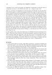
Purchased for the exclusive use of nofirst nolast (unknown)
From: SCC Media Library & Resource Center (library.scconline.org)